
Page 1914 - Hematology_ Basic Principles and Practice ( PDFDrive )
P. 1914
1694 Part XI Transfusion Medicine
Blood Group Systems add specific monosaccharides in specific linkages to a growing oligo-
15
saccharide precursor chain (reviewed in Clausen and Hakomori ).
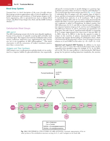
Presented here is a brief description of the most clinically relevant The terminal sugar determines antigen specificity (Fig. 110.2). Group
blood group systems in approximate order of clinical significance. For O individuals have H antigen only, the terminal sugar of which is
further information and prevalence of blood group antigens in dif- fucose, and this is the precursor substrate for A and B antigens. Group
ferent populations, refer to specialized texts such as Human Blood O individuals have defective A or B transferases. The A and B
Groups, The Blood Group Antigen Facts Book, and the AABB Technical transferase enzymes differ only by the nature of the monosaccharide
Manual. 2,3,14 added to the chain. N-acetyl-D-galactosamine is added by A-transferase,
and D-galactose is added by B-transferase. In clinical practice, four
ABO phenotypes (A, B, O, and AB) are discriminated. In addition,
two common variations of group A (A 1 and A 2) can be distinguished.
Carbohydrate Blood Groups The differences between A 1 and A 2 phenotypes are quantitative and
qualitative. Not only is the A 1 transferase more efficient in converting
ABO and H H to A antigen (approximately five times more A sites per RBC on
The ABO blood group system is by far the most clinically significant, A 1 RBCs than on A 2 RBCs), it also has the capacity to make A 1
because of the presence of naturally occurring IgM antibodies (and antigen on the repetitive A epitope. Quantitatively normal ABH
sometimes IgG). The original observation by Landsteiner that certain expression also requires the branching of carbohydrate chains, which
human erythrocyte suspensions were agglutinated by other human is performed by the blood group I enzyme. Some H antigen precursor
14a
sera led to the recognition of ABO polymorphism. This initial remains on A and B RBCs in this order: A 2 > B > A 2B > A 1 > A 1 B.
observation is still the cornerstone of modern transfusion practice
more than a century later. Inherited and Acquired ABH Variation In addition to the main
ABO types, there are many other inherited phenotypes with a weaker
Antigens and Their Synthesis expression of the specified antigen, for example, A 3 , A x , A el , B 3, B(A),
ABH antigens occur on glycoproteins and glycolipids and are synthe- and cis-AB. This can cause problems in determining the ABO blood
sized in a stepwise fashion by glycosyltransferases that sequentially group, but for patients needing immediate transfusion, the selection
Gal GlcNAc R
Precursor: β1 4
Gal GlcNAc
β1 4 β1 3
Fucosyltransferase H (FUTI)
Gene
Gal GlcNAc R
β1 4
H Gal GlcNAc
β1 4 β1 3
α1 2
Fuc
A transferase B transferase
A B
Gal GlcNAc R Gal GlcNAc R
β1 4 β1 4
GalNAc Gal GlcNAc Gal Gal GlcNAc
α1 4 β1 4 β1 3 β1 4 β1 3
α1 2 α1 2
Fuc Fuc
Gal = Galactose
Fuc = Fucose
GalNAc = N-acetylgalactosamine
GlcNAc = N-acetylglucosamine
Fig. 110.2 BIOCHEMICAL STRUCTURES OF ABH ANTIGENS. Schematic representation of the ter-
minal portions of the carbohydrate structures carrying the H, A, and B antigens on red blood cells.