
Page 1916 - Hematology_ Basic Principles and Practice ( PDFDrive )
P. 1916
1696 Part XI Transfusion Medicine
normal state on RBCs from fetuses and infants. The gene encoding C or c and E or e antigens carried with D are represented by sub-
the I-branching β-1,6-N-acetylglucosaminyltransferase (GCNT2) has scripts: 1 for Ce (R 1), 2 for cE (R 2), 0 for ce (R 0), and Z for CE (R z ).
three alternative forms of exon 1 with common exons 2 and 3. The presence of these antigens without D is represented by a super-
y
Mutations in exon 2 or exon 3 silence GCNT2 and give rise to the script: prime for Ce (r′), double-prime for cE (r″), and y for CE (r ).
form of the i phenotype that is associated with cataracts in Asians. This terminology allows one to convey the common Rh antigens (the
Mutations in exon 1C silence the gene in erythrocytes (but not in phenotype) with a single term. The third system of numeric designa-
other tissues) and lead to the i phenotype without cataracts. tions is not widely used in the laboratory, with a few exceptions
Alloanti-I made by a person with the rare i adult phenotype can (Rh17, Rh32, Rh33).
be clinically significant and cause destruction of transfused I-positive
RBCs. However, the sera of all I-positive individuals contain autoanti-I Genes, Proteins, Antigens, and Phenotypes The Rh proteins are
that is clinically benign and reactive only at or below room tempera- designated RhD (encoded by RHD), which carries the D antigen,
ture. In contrast, cold hemagglutinin disease is characterized by a high and RhCE (encoded by RHCE), which carries the CE antigens
titer of complement-fixing monoclonal anti-I, which causes in vivo (either ce, cE, Ce, or CE). RhD differs from the various forms of
hemolysis and hemolytic anemia. The titer and thermal range of RhCE by 32–35 amino acids. RhD and RhCE are not glycosylated
autoanti-I is often increased following infection with Mycoplasma but form a complex in the RBC membrane with RhAG (Rh-associated
pneumoniae. If transfusion cannot be avoided, donor RBCs should glycoprotein). Other proteins present in the Rh-complex are CD47
be transfused through a blood warmer. (an integrin-associated protein), LW, and glycophorin B. The
The FORS1 blood group antigen is a rare low prevalence antigen Rh-complex also associates with band 3 (the anion exchanger) as a
that is A-like in that it is defined by a terminal N-acetylgalactosamine macrocomplex in the membrane.
and was first recognized as a weak A subgroup. The antigen has been The D-negative (Rh-negative) phenotype is prevalent in whites
defined as Forssman antigen, commonly found on the RBCs of (15%–17%), less common in African blacks (3%–5%), and rare in
nonprimate mammals, and arises from a gain-of-function mutation Asians (<0.1%). The absence of D in Europeans is primarily caused
in the GBGT1 pseudogene. The clinical relevance of anti-FORS1 by a deletion of the RHD gene. African blacks and rare D-negative
found naturally occurring in the plasma of most individuals is not whites and Asians carry a RHD gene that is silenced by a variety of
known. molecular events. 3
RBCs with weak D have D antigen but at lower levels than normal
because of one or more amino acid changes that are often predicted to
be in the intracellular or transmembrane regions of RhD. The RBCs
Protein Blood Groups do not lack, or have altered, epitopes of D. Many individuals with a
serologic weak D phenotype have weak D types 1, 2, and 3 by RHD
Rhe, Rhe-associated glycoprotein, and genotyping, and individuals with these genotypes can safely receive
LW Blood Group Systems D-positive blood and do not make clinically significant anti-D. 21,22
The Rh system is second only to the ABO system in importance in Partial D antigens (previously called D categories or D mosaics) are
transfusion medicine. Rh antigens, especially D, are highly immuno- caused either by point mutations in RHD that encode amino acid
genic; thus in most countries, blood for transfusion is tested and changes that alter D epitopes, or by replacement of RHD nucleotides
labeled with the D antigen type (Rh-positive or Rh-negative) and or exons by the equivalent part of RHCE that result in loss of D
D−recipients are transfused with D−RBC products. epitopes. RBCs with a partial D antigen may have strong or weak
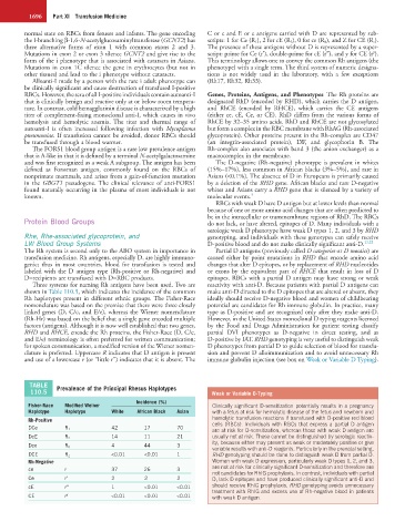
Three systems for naming Rh antigens have been used. Two are reactivity with anti-D. Because patients with partial D antigens can
shown in Table 110.5, which indicates the incidence of the common make anti-D directed to the D epitopes that are altered or absent, they
Rh haplotypes present in different ethnic groups. The Fisher-Race ideally should receive D-negative blood and women of childbearing
nomenclature was based on the premise that there were three closely potential are candidates for Rh immune globulin. In practice, many
linked genes (D, C/c, and E/e), whereas the Wiener nomenclature type as D-positive and are recognized only after they make anti-D.
(Rh-Hr) was based on the belief that a single gene encoded multiple However, in the United States monoclonal D typing reagents licensed
factors (antigens). Although it is now well established that two genes, by the Food and Drugs Administration for patient testing classify
RHD and RHCE, encode the Rh proteins, the Fisher-Race (D, C/c, partial DVI phenotypes as D-negative in direct testing, and as
and E/e) terminology is often preferred for written communication; D-positive by IAT. RHD genotyping is very useful to distinguish weak
for spoken communication, a modified version of the Wiener nomen- D phenotypes from partial D to guide selection of blood for transfu-
clature is preferred. Uppercase R indicates that D antigen is present sion and prevent D alloimmunization and to avoid unnecessary Rh
and use of a lowercase r (or “little r”) indicates that it is absent. The immune globulin injection (see box on Weak or Variable D Typing).
TABLE Prevalence of the Principal Rhesus Haplotypes
110.5 Weak or Variable D-Typing
Incidence (%)
Fisher-Race Modified Weiner Clinically significant D-sensitization potentially results in a pregnancy
Haplotype Haplotype White African Black Asian with a fetus at risk for hemolytic disease of the fetus and newborn and
Rh-Positive hemolytic transfusion reactions if transfused with D-positive red blood
cells (RBCs). Individuals with RBCs that express a partial D antigen
DCe R 1 42 17 70 are at risk for D-sensitization, whereas those with weak D antigen are
DcE R 2 14 11 21 usually not at risk. These cannot be distinguished by serologic reactiv-
ity, because either may present as weak or moderately positive or give
Dce R 0 4 44 3
variable results with anti-D reagents. Particularly in the prenatal setting,
DCE R Z <0.01 <0.01 1 RHD genotyping should be done to distinguish weak D from partial D.
Rh-Negative Women with weak D expression, particularly weak D types 1, 2, and 3,
ce r 37 26 3 are not at risk for clinically significant D-sensitization and therefore are
not candidates for RhIG prophylaxis. In contrast, individuals with partial
Ce r′ 2 2 2 D, lack D epitopes and have produced clinically significant anti-D and
cE r″ 1 <0.01 <0.01 should receive RhIG prophylaxis. RHD genotyping avoids unnecessary
treatment with RhIG and excess use of Rh-negative blood in patients
CE r y <0.01 <0.01 <0.01 with weak D antigen.