
Page 2116 - Hematology_ Basic Principles and Practice ( PDFDrive )
P. 2116
Chapter 125 Molecular Basis of Platelet Function 1879
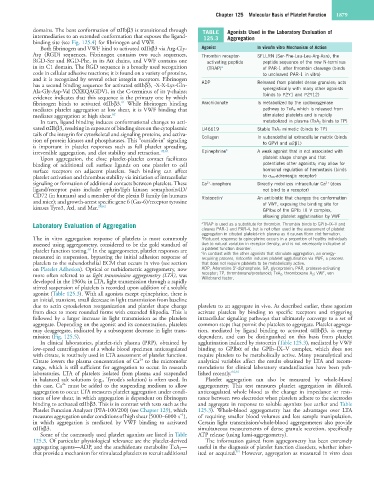
domains. The bent conformation of αIIbβ3 is transitioned through TABLE Agonists Used in the Laboratory Evaluation of
intermediaries to an extended conformation that exposes the ligand- 125.3 Aggregation
binding site (see Fig. 125.4) for fibrinogen and VWF.
Both fibrinogen and VWF bind to activated αIIbβ3 via Arg-Gly- Agonist In vivo/In vitro Mechanism of Action
Asp (RGD) sequences. Fibrinogen contains two such sequences, Thrombin receptor- SFLLRN (Ser-Phe-Leu-Leu-Arg-Asn), the
RGD-Ser and RGD-Phe, in its Aα chains, and VWF contains one activating peptide peptide sequence of the new N-terminus
in its C1 domain. The RGD sequence is a broadly used recognition (TRAP) a of PAR-1 after thrombin cleavage (binds
code in cellular adhesive reactions; it is found on a variety of proteins, to uncleaved PAR-1 in vitro)
and it is recognized by several other integrin receptors. Fibrinogen
has a second binding sequence for activated αIIbβ3, -X-X-Lys-Gln- ADP Released from platelet dense granules; acts
Ala-Gly-Asp-Val (XXKQAGDV), in the C-terminus of its γ-chains; synergistically with many other agonists
evidence indicates that this sequence is the primary one by which (binds to P2Y1 and P2Y12)
61
fibrinogen binds to activated αIIbβ3. While fibrinogen binding Arachidonate Is metabolized by the cyclooxygenase
mediates platelet aggregation at low shear, it is VWF binding that pathway to TxA 2 which is released from
mediates aggregation at high shear. 62 stimulated platelets and is rapidly
In turn, ligand binding induces conformational changes to acti- metabolized in plasma (TxA 2 binds to TP)
vated αIIbβ3, resulting in exposure of binding sites on the cytoplasmic U46619 Stable TxA 2 mimetic (binds to TP)
tails of the integrin for cytoskeletal and signaling proteins, and activa- Collagen In subendothelial extracellular matrix (binds
tion of protein kinases and phosphatases. This “outside-in” signaling to GPVI and α2β1)
is important in platelet responses such as full platelet spreading,
irreversible aggregation, and clot stability and retraction. 28,61 Epinephrine b A weak agonist that is not associated with
Upon aggregation, the close platelet-platelet contact facilitates platelet shape change and that
binding of additional cell surface ligands on one platelet to cell potentiates other agonists; may allow for
surface receptors on adjacent platelets. Such binding can affect hormonal regulation of hemostasis (binds
platelet activation and thrombus stability via initiation of intracellular to α 2A -adrenergic receptor)
signaling or formation of additional contacts between platelets. These Ca -ionophore Directly mobilizes intracellular Ca (does
2+
2+
ligand/receptor pairs include: ephrin/Eph kinase; semaphorin4D/ not bind to a receptor)
CD72 (in humans) and a member of the plexin B family (in humans Ristocetin c An antibiotic that changes the conformation
and mice); and growth-arrest specific gene 6 (Gas-6)/receptor tyrosine of VWF, exposing the binding site for
kinases Tyro3, Axl, and Mer. 28,62 GPIbα of the GPIb–IX–V complex,
allowing platelet agglutination by VWF
Laboratory Evaluation of Aggregation a TRAP is used as a substitute for thrombin. Thrombin binds to GPIb–IX–V and
cleaves PAR-1 and PAR-4, but is not often used in the assessment of platelet
aggregation in citrated platelet-rich plasma as it causes fibrin clot formation.
The in vitro aggregation response of platelets is most commonly b Reduced response to epinephrine occurs in a proportion of healthy individuals
assessed using aggregometry, considered to be the gold standard of due to natural variation in receptor density, and is not necessarily indicative of
63
platelet function testing. In the aggregometer, platelet responses are a platelet function disorder.
In contrast with the other agonists that stimulate aggregation, an energy-
c
measured in suspension, bypassing the initial adhesion response of requiring process, ristocetin induces platelet agglutination via VWF, a process
platelets to the subendothelial ECM that occurs in vivo (see section that does not require platelets to be metabolically active.
on Platelet Adhesion). Optical or turbidometric aggregometry, now ADP, Adenosine 5′-diphosphate; GP, glycoprotein; PAR, protease-activating
more often referred to as light transmission aggregometry (LTA), was receptor; TP, thromboxane/prostanoid; TxA 2 , thromboxane A 2 ; VWF, von
Willebrand factor.
developed in the 1960s; in LTA, light transmission through a rapidly
stirred suspension of platelets is recorded upon addition of a soluble
agonist (Table 125.3). With all agonists except epinephrine, there is
an initial, transient, small decrease in light transmission from baseline
due to actin cytoskeleton reorganization and platelet shape change platelets to an aggregate in vivo. As described earlier, these agonists
from discs to more rounded forms with extended filipodia. This is activate platelets by binding to specific receptors and triggering
followed by a larger increase in light transmission as the platelets intracellular signaling pathways that ultimately converge to a set of
aggregate. Depending on the agonist and its concentration, platelets common steps that permit the platelets to aggregate. Platelet aggrega-
may deaggregate, indicated by a subsequent decrease in light trans- tion, mediated by ligand binding to activated αIIbβ3, is energy
mission (Fig. 125.5). dependent, and can be distinguished on this basis from platelet
In clinical laboratories, platelet-rich plasma (PRP), obtained by agglutination induced by ristocetin (Table 125.3), mediated by VWF
low-speed centrifugation of a whole blood specimen anticoagulated binding to GPIbα of the GPIb–IX–V complex, which does not
with citrate, is routinely used in LTA assessment of platelet function. require platelets to be metabolically active. Many preanalytical and
2+
Citrate lowers the plasma concentration of Ca to the micromolar analytical variables affect the results obtained by LTA and recom-
range, which is still sufficient for aggregation to occur. In research mendations for clinical laboratory standardization have been pub-
laboratories, LTA of platelets isolated from plasma and suspended lished recently. 64,65
in balanced salt solutions (e.g., Tyrode’s solution) is often used. In Platelet aggregation can also be measured by whole-blood
2+
this case, Ca must be added to the suspending medium to allow aggregometry. This test measures platelet aggregation in diluted,
aggregation to occur. LTA measures platelet aggregation under condi- anticoagulated whole blood as the change in impedance or resis-
tions of low shear, in which aggregation is dependent on fibrinogen tance between two electrodes when platelets adhere to the electrodes
binding to activated αIIbβ3. This is in contrast with tests such as the and aggregate in response to soluble agonists (see earlier and Table
Platelet Function Analyzer (PFA-100/200) (see Chapter 129), which 125.3). Whole-blood aggregometry has the advantages over LTA
−1
measures aggregation under conditions of high shear (5000–6000 s ), of requiring smaller blood volumes and less sample manipulation.
in which aggregation is mediated by VWF binding to activated Certain light transmission/whole-blood aggregometers also provide
αIIbβ3. simultaneous measurements of dense granule secretion, specifically
Some of the commonly used platelet agonists are listed in Table ATP release (using lumi-aggregometry).
125.3. Of particular physiological relevance are the platelet-derived The information gained from aggregometry has been extremely
aggregating agents—ADP, and the arachidonate metabolite TxA 2 — useful in the diagnosis of platelet function disorders, whether inher-
64
that provide a mechanism for stimulated platelets to recruit additional ited or acquired. However, aggregation as measured in vitro does