
Page 2115 - Hematology_ Basic Principles and Practice ( PDFDrive )
P. 2115
1878 Part XII Hemostasis and Thrombosis
outer cytoplasmic leaflet, and the minor anionic aminophospholipid Ligand
PS predominating in the inner. Platelet adhesion to collagen and
stimulation with thrombin synergistically result in a loss of the asym-
metry, or “scrambling,” of the membrane phospholipids, such that
53
PS becomes exposed on the external plasma membrane leaflet. PS
translocated to the surface of activated platelets facilitates the assem-
bly of the intrinsic tenase complex (factors VIIIa, IXa, and X) and
prothrombinase complex (factors Va and Xa, and prothrombin),
contributing to the burst of thrombin generation that occurs in the
propagation phase of coagulation (see Chapter 126). Specifically, the
negatively charged γ-carboxyglutamate (Gla) residues of the vitamin
K-dependent factors, factors VII(a), IX(a), X(a), and prothrombin,
2+
mediate Ca -dependent binding of the factors to negatively-charged Membrane
PS. There is recent evidence that plasminogen also binds to IIb IIb 3
PS-exposing platelets, thereby linking coagulation and fibrinolysis. 54 3 Talin-H
Exposure of PS on the platelet surface requires sustained increases Cytoplasm
2+
in [Ca ] i via influx across the plasma membrane, and activation of Kindlin
phospholipid scramblase (see Chapter 130, Fig. 130.1), which rapidly
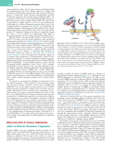
and nonspecifically transports phospholipids between the membrane Fig. 125.4 MODEL SHOWING THE ACTIVATION OF αIIbβ3. Signal-
leaflets. The transmembrane protein TMEM16F (anoctamin 6) has ing pathways induced by platelet agonists result in talin and kindlin binding
55
recently been shown to be essential for PS exposure, but its specific to the cytoplasmic tail of β3 and inside-out signaling such that the integrin
role is not yet clear. Scott syndrome, a platelet disorder associated is transformed from its low-affinity bent conformation on the resting platelet
with a defect in PS exposure, is associated with mutations in to its high-affinity extended conformation on the activated platelet. The
TMEM16F (ANO6; see section on Molecular Basis of Inherited extended conformation has the ligand binding site exposed, allowing for the
Platelet Disorders). There is heterogeneity in the platelet response to binding of fibrinogen or von Willebrand factor, and thus for platelet aggrega-
stimulation: only a proportion of activated platelets expose PS (this tion. Binding of these ligands initiates outside-in signaling, triggering further
population of platelets is variously described as being procoagulant, intracellular signal transduction and platelet responses. This model is based
superactivated, coated, or having Sustained Calcium-Induced Platelet on the crystal structure of the extracellular domain of αvβ3, and the cyto-
56
[SCIP] morphology). Coated platelets appear to overlap with the plasmic tails on the nuclear magnetic resonance structure of αIIbβ3. (Adapted,
PS-exposing platelet population, being “coated” with covalently with permission, from Bledzka K, Smyth SS, Plow EF. Integrin αIIbβ3. From discov-
linked platelet secretion products (e.g., serotonin, fibrinogen, factor ery to efficacious therapeutic target. Circ Res 112:1189, 2013.)
Va). Besides supporting thrombin generation, PS-exposing platelets
are characterized by inactive αIIbβ3 and a distinct balloon-like
morphology with loss of internal organelles. In vitro, they are readily
identified by annexin A5 or lactadherin binding. The pathway that to bind its ligands. Activation of αIIbβ3 results in a change to a
leads to rapid PS exposure on activated platelets can be distinguished high-affinity, extended conformation (Fig. 125.4), allowing it to bind
from a slower intrinsic apoptotic pathway, which involves caspase divalent fibrinogen or multivalent VWF which can bridge to other
activation. 57 activated αIIbβ3 molecules on adjacent platelets, mediating platelet
PS exposure is accompanied by blebbing of microparticles from aggregation. The N-terminal portion of β3 contains three divalent
58
the plasma membrane. These small (0.1–1 µm diameter) vesicles cation-binding sites including the metal-ion dependent adhesion site
2+
not only express platelet membrane receptors such as αIIbβ3 and (MIDAS); metal ions (Ca ) are required for ligand binding to
GPIb–IX–V, but can also express activation markers such as CD62P. αIIbβ3. Absence or dysfunction of αIIbβ3 on the platelet surface
Platelet-derived microparticles are distinct from exosomes, which are leads to Glanzmann thrombasthenia (GT), a disorder characterized
smaller (40–100 nm) and are released from intracellular vesicles. by a moderate-to-severe bleeding diathesis and absent in vitro platelet
Microparticles can support hemostasis; platelets from patients with aggregation responses to all agonists (see section on Molecular Basis
the very rare bleeding disorder Castaman syndrome cannot generate of Inherited Platelet Disorders).
microparticles. Circulating microparticles are considered to originate Activation of platelets with any of the agonists described in the
from megakaryocytes as well as from platelets. Increased circulating preceding sections, including collagen, thrombin, ADP, and TxA 2 ,
levels of microparticles have been reported in thrombotic conditions results in the conversion of αIIbβ3 from its resting to its activated
60
such as myocardial infarction, thrombotic thrombocytopenic purpura, state. This transformation is a consequence of the intracellular sig-
sickle cell disease, and diabetes, immune-mediated conditions such naling pathways set in motion when the agonist binds to its receptor
as immune thrombocytopenia and heparin-induced thrombocytope- (see section on Activation Pathways), leading to the final common
nia, and in malignancy and inflammatory conditions. Microparticles pathway that results in aggregation; it occurs rapidly, on the order of
formed by platelets activated via GPVI have been shown to contribute seconds, after platelet exposure to an agonist. The signal is transmit-
to the pathophysiology of rheumatoid arthritis, delivering IL-1 to ted to the cytoplasmic tails of αIIbβ3 and from the cytoplasmic tail
59
synovial fibroblasts, an example of how microparticles may play a through the transmembrane helices, ultimately inducing a change
role in platelet-cell communication by delivering bioactive molecules in the extracellular domain to render αIIbβ3 competent to bind its
to target cells. ligands. The signaling process responsible for this transformation is
referred to as “inside-out” signaling through αIIbβ3.
2+
Specifically, it is the agonist-induced increase in [Ca ] i and DAG
MOLECULAR BASIS OF PLATELET AGGREGATION formation that result in the final common pathway of integrin activa-
2+
61
tion. Via the Ca - and DAG-regulated guanine nucleotide exchange
αIIbβ3 and Molecular Mechanisms of Aggregation factor (CalDAG-GEFI), the small monomeric G protein (GTPase)
Rap1 is activated and binds to Rap1-GTP-interacting adaptor mol-
Integrin αIIbβ3 is the most abundant membrane protein on the ecule (RIAM); RIAM promotes binding of the cytoskeletal protein
platelet surface, with 40,000–80,000 copies per resting platelet, a very talin to the cytoplasmic domain of β3. Talin cooperates with kindlin-
high receptor density for such a small cell. Platelet activation can 3, which also binds to the cytoplasmic domain of β3, to unclasp the
produce an additional 10% increase in this number as a result of complex between the αIIb and β3 cytoplasmic tails. This leads to a
expression from internal pools, i.e., α-granule membranes. On resting dissociation of the transmembrane complex of the αIIb and β3
platelets, αIIbβ3 is in a low-affinity bent conformation that is unable subunits, inducing a conformational change in the extracellular