
Page 2174 - Hematology_ Basic Principles and Practice ( PDFDrive )
P. 2174
Chapter 129 Laboratory Evaluation of Hemostatic and Thrombotic Disorders 1927
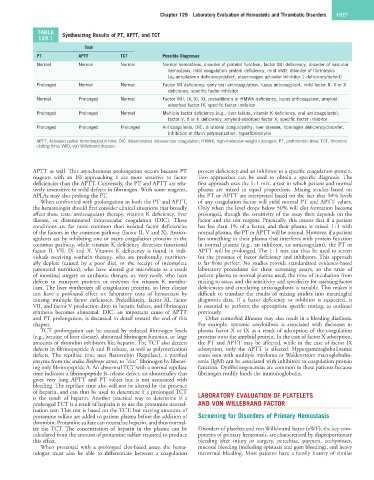
TABLE Synthesizing Results of PT, APTT, and TCT
129.1
Test
PT APTT TCT Possible Diagnoses
Normal Normal Normal Normal hemostasis, disorder of platelet function, factor XIII deficiency, disorder of vascular
hemostasis, mild coagulation protein deficiency, mild vWD, disorder of fibrinolysis
(α 2 -antiplasmin deficiency/defect, plasminogen activator inhibitor-1 deficiency/defect)
Prolonged Normal Normal Factor VII deficiency, early oral anticoagulation, lupus anticoagulant, mild factor II, V or X
deficiency, specific factor inhibitor
Normal Prolonged Normal Factor VIII, IX, XI, XI, prekallikrein or HMWK deficiency, lupus anticoagulant, amyloid-
adsorbed factor IX, specific factor inhibitor
Prolonged Prolonged Normal Multiple factor deficiency (e.g., liver failure, vitamin K deficiency, oral anticoagulants),
factor V, X or II deficiency, amyloid-adsorbed factor X, specific factor inhibitor
Prolonged Prolonged Prolonged Anticoagulants, DIC, dilutional coagulopathy, liver disease, fibrinogen deficiency/disorder,
inhibition of fibrin polymerization, hyperfibrinolysis
APTT, Activated partial thromboplastin time; DIC, disseminated intravascular coagulation; HMWK, high-molecular-weight kininogen; PT, prothrombin time; TCT, thrombin
clotting time; vWD, von Willebrand disease.
APTT as well. This asynchronous prolongation occurs because PT protein deficiency and an inhibitor to a specific coagulation protein.
reagents with an ISI approaching 1 are more sensitive to factor Two approaches can be used to obtain a specific diagnosis. The
deficiencies than the APTT. Conversely, the PT and APTT are rela- first approach uses the 1 : 1 mix, a test in which patient and normal
tively insensitive to mild defects in fibrinogen. With some reagents, plasma are mixed in equal proportions. Mixing studies based on
APLAs may also prolong the PT. the PT or APTT are interpreted based on the fact that 50% levels
When confronted with prolongation in both the PT and APTT, of any coagulation factor will yield normal PT and APTT values.
the hematologist should first consider clinical situations that broadly Only when the level drops below 50% will clot formation become
affect these tests: anticoagulant therapy, vitamin K deficiency, liver prolonged, though the sensitivity of the assay then depends on the
disease, or disseminated intravascular coagulation (DIC). These factor and the test reagent. Practically, this means that if a patient
conditions are far more common than isolated factor deficiencies has less than 1% of a factor, and their plasma is mixed 1 : 1 with
of the factors in the common pathway (factor II, V and X). Antico- normal plasma, the PT or APTT will be normal. However, if a patient
agulants act by inhibiting one or many coagulation proteins in the has something in their plasma that interferes with protein function
common pathway, while vitamin K deficiency decreases functional in normal plasma (e.g., an inhibitor, an anticoagulant), the PT or
factor II, VII, IX and X. Vitamin K deficiency is found in indi- APTT will be prolonged. The 1 : 1 mix can thus be used to screen
viduals receiving warfarin therapy, who are profoundly nutrition- for the presence of factor deficiency and inhibitors. This approach
ally deplete (caused by a poor diet, or the receipt of incomplete is far from perfect. No studies provide standardized evidence-based
parenteral nutrition), who have altered gut microbiota as a result laboratory procedures for these screening assays, so the ratio of
of intestinal surgery or antibiotic therapy, or, very rarely, who have patient plasma to normal plasma used, the time of incubation from
defects in transport proteins or enzymes for vitamin K metabo- mixing to assay, and the sensitivity and specificity for assessing factor
lism. The liver synthesizes all coagulation proteins, so liver disease deficiencies and circulating anticoagulants is variable. This makes it
can have a profound effect on laboratory tests of hemostasis by difficult to translate the results of mixing studies into meaningful
causing multiple factor deficiency. Prekallikrein, factor XI, factor diagnostic data. If a factor deficiency or inhibitor is suspected, it
VII, and factor V production drop in hepatic failure, and fibrinogen is essential to perform the appropriate specific testing, as outlined
synthesis becomes abnormal. DIC, an important cause of APTT previously.
and PT prolongation, is discussed in detail toward the end of this Other comorbid illnesses may also result in a bleeding diathesis.
chapter. For example, systemic amyloidosis is associated with decreases in
TCT prolongation can be caused by reduced fibrinogen levels plasma factor X or IX as a result of adsorption of the coagulation
(e.g., because of liver disease), abnormal fibrinogen function, or large proteins onto the amyloid protein. In the case of factor X adsorption,
amounts of thrombin inhibitors like heparin. The TCT also detects the PT and APTT may be affected, while in the case of factor IX
defects in fibrinopeptide A and B release, as well as polymerization adsorption, only the APTT is affected. Hypergammaglobulinemic
defects. The reptilase time uses Batroxobin (Reptilase), a purified states seen with multiple myeloma or Waldenström macroglobulin-
enzyme from the snake Bothrops atrox, to “clot” fibrinogen by liberat- emia (IgM) can be associated with inhibitors to coagulation protein
ing only fibrinopeptide A. An abnormal TCT with a normal reptilase function. Dysfibrinogenemias are common in these patients because
time indicates a fibrinopeptide B–release defect, an abnormality that fibrinogen readily binds the immunoglobulin.
gives very long APTT and PT values but is not associated with
bleeding. The reptilase time also will not be altered by the presence
of heparin, and can thus be used to determine if a prolonged TCT
is the result of heparin. Another practical way to determine if a LABORATORY EVALUATION OF PLATELETS
prolonged TCT is a result of heparin is to use the protamine neutral- AND VON WILLEBRAND FACTOR
ization test. This test is based on the TCT, but varying amounts of
protamine sulfate are added to patient plasma before the addition of Screening for Disorders of Primary Hemostasis
thrombin. Protamine sulfate can neutralize heparin, and thus normal-
ize the TCT. The concentration of heparin in the plasma can be Disorders of platelets and von Willebrand factor (vWF), the key com-
calculated from the amount of protamine sulfate required to produce ponents of primary hemostasis, are characterized by disproportionate
this effect. bleeding after injury or surgery, petechiae, purpura, ecchymoses,
When presented with a prolonged clot-based assay, the hema- mucosal bleeding (including epistaxis and gum bleeding), and heavy
tologist must also be able to differentiate between a coagulation menstrual bleeding. Most patients have a family history of similar