
Page 2247 - Hematology_ Basic Principles and Practice ( PDFDrive )
P. 2247
1994 Part XII Hemostasis and Thrombosis
Protease except erythrocytes. Mutations are found throughout the extracellular
C3 C3b + C3a domain of the protein (see Fig. 134.7) and most commonly lead to
A diminished cell surface expression, although some impair protein
activity.
C3 MCP and factor H bind C3b and facilitate its cleavage on the cell
membrane by factor I. Factor I mutations are observed in approxi-
mately 12% of aHUS patients and most commonly result in decreased
protein expression; although some mutations cause decreased catalytic
AP
activity, which is mediated through the factor I light chain. Throm-
B, D, P bomodulin also enhances CFI-mediated degradation of C3b, and
C3b mutations of thrombomodulin have been observed in 5% of patients
with aHUS in one series.
B, SP C3b + B → C3bB Mutations in factor B and C3 are observed in approximately 3%
D, SP C3bB + D → C3bBb and 10% of aHUS patients, respectively. Mutations in factor B lead
B P, stabilizer C3bBb + P → C3bBbP to enhanced formation or greater stability of C3 convertase on cell
surfaces. Mutations in C3 may result in resistance to regulation,
principally mediated by diminished ability of regulatory proteins
Alternative pathway (CHF, MCP or CFI) to interact with mutant C3b.
C3 convertase Recently, mutations in the gene encoding for DGKE, a lipid
C3 C3bBbP kinase expressed in endothelium, platelets, and renal podocytes,
has been identified as the cause of an autosomal recessive form of
aHUS with high penetrance. The mechanism of disease is attributed
to deregulation of intracellular signaling, leading to activation of
AP AP protein kinase C with a subsequent shift of the balance of endothe-
lial cells and platelets toward a more activated and prothrombotic
B, D, P phenotype. Altered podocyte homeostasis has been hypothesized to
C3b occur as a consequence of abnormal protein kinase C-dependent
Endothelial cell C3a and C5a vascular endothelial growth factor (VEGF) receptor expression,
membrane disrupting this important cell nurturing pathway. In general,
(Anaphylatoxins) patients with DGKE-related aHUS have no evidence of comple-
C C5b-C9 ment dysregulation. Furthermore, unlike patients with defects of
the complement mechanism, patients with DGKE-associated aHUS
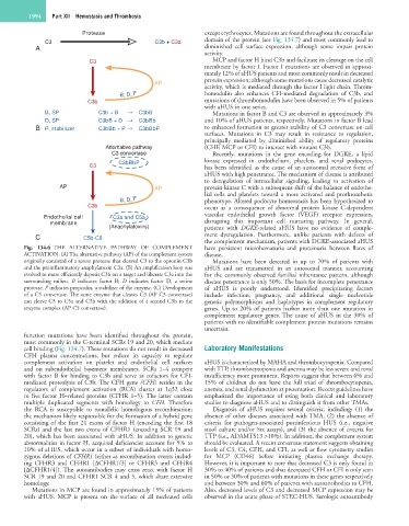
Fig. 134.6 THE ALTERNATIVE PATHWAY OF COMPLEMENT have persistent microhematuria and proteinuria between flares of
ACTIVATION. (A) The alternative pathway (AP) of the complement system disease.
originally consisted of a serine protease that cleaved C3 to the opsonin C3b Mutations have been detected in up to 70% of patients with
and the proinflammatory anaphylatoxin C3a. (B) An amplification loop was aHUS and are transmitted in an autosomal manner, accounting
evolved to more efficiently deposit C3b on a target and liberate C3a into the for the commonly observed familial inheritance pattern, although
surrounding milieu. B indicates factor B; D indicates factor D, a serine disease penetrance is only 50%. The basis for incomplete penetrance
protease; P indicates properdin, a stabilizer of the enzyme. (C) Development of aHUS is poorly understood. Identified precipitating factors
of a C5 convertase. The same enzyme that cleaves C3 (AP C3 convertase) include infection, pregnancy, and additional single nucleotide
can cleave C5 to C5a and C5b with the addition of a second C3b to the genetic polymorphisms and haplotypes in complement regulatory
enzyme complex (AP C5 convertase). genes. Up to 20% of patients harbor more than one mutation in
complement regulatory genes. The cause of aHUS in the 30% of
patients with no identifiable complement protein mutations remains
uncertain.
function mutations have been identified throughout the protein,
most commonly in the C-terminal SCRs 19 and 20, which mediate
cell binding (Fig. 134.7). These mutations do not result in decreased Laboratory Manifestations
CFH plasma concentrations, but reduce its capacity to regulate
complement activation on platelet and endothelial cell surfaces aHUS is characterized by MAHA and thrombocytopenia. Compared
and on subendothelial basement membranes. SCRs 1–4 compete with TTP, thrombocytopenia and anemia may be less severe and renal
with factor B for binding to C3b and serve as cofactors for CFI- insufficiency more prominent. Reports suggest that between 6% and
mediated proteolysis of C3b. The CFH gene (CFH) resides in the 15% of children do not have the full triad of thrombocytopenia,
regulators of complement activation (RCA) cluster at 1q32 close anemia, and renal dysfunction at presentation. Recent guidelines have
to five factor H–related proteins (CFHR 1–5). The latter contain emphasized the importance of using both clinical and laboratory
multiple duplicated segments with homology to CFH. Therefore studies to diagnose aHUS and to distinguish it from other TMAs.
the RCA is susceptible to nonallelic homologous recombination; Diagnosis of aHUS requires several criteria, including: (1) the
the mechanism likely responsible for the formation of a hybrid gene absence of other diseases associated with TMA, (2) the absence of
consisting of the first 21 exons of factor H (encoding the first 18 criteria for pathogen-associated postinfectious HUS (i.e., negative
SCRs) and the last two exons of CFHR1 (encoding SCR 19 and stool culture and/or Stx assays), and (3) the absence of criteria for
20), which has been associated with aHUS. In addition to genetic TTP (i.e., ADAMTS13 >10%). In addition, the complement system
abnormalities in factor H, acquired deficiencies account for 5% to should be evaluated. A recent consensus statement suggests obtaining
10% of aHUS, which occur in a subset of individuals with homo- levels of C3, C4, CFH, and CFI, as well as flow cytometry studies
zygous deletions of CFHR1 (either as recombination events includ- for MCP (CD46) before initiating plasma exchange therapy.
ing CFHR3 and CFHR1 [ΔCFHR1/3] or CFHR3 and CFHR4 However, it is important to note that decreased C3 is only found in
[ΔCFHR1/4]). The autoantibodies may cross react with factor H 30% to 40% of patients and that decreased CFH or CFI is only seen
SCR 19 and 20 and CFHR1 SCR 4 and 5, which share extensive in 50% or 30% of patients with mutations in these genes respectively
homology. and between 30% and 60% of patients with autoantibodies to CFH.
Mutations in MCP are found in approximately 15% of patients Also, decreased levels of C3 and decreased MCP expression may be
with aHUS. MCP is present on the surface of all nucleated cells observed in the acute phase of STEC-HUS. Serologic autoantibody