
Page 228 - Hematology_ Basic Principles and Practice ( PDFDrive )
P. 228
180 Part II Cellular Basis of Hematology
D-CDK4/6 can sequester p21, preventing inactivation of CDK2- is its upregulation in aging tissues and in response to oncogene activa-
containing complexes. tion. For example, in response to continuously high RAS or E2F
In marked contrast with the transcriptional regulation of p21, activity, CDKN2A is transcriptionally induced, leading to cell cycle
regulation of p27 (KIP1, CDKN1B) occurs posttranscriptionally, arrest and senescence.
such that mRNA levels remain largely constant, whereas levels of the
protein change. p27 protein levels are maximal during quiescence and
early G 1, and p27 protein binds and inhibits cyclin E–CDK2 com- TRANSCRIPTIONAL REGULATION: MYC,
plexes. The progressive decrease in p27 protein levels during G 1 RB-E2F, DREAM, AND MMB-FOXM1
allows for activation of cyclin E–CDK2 complexes that are required
for the transition into S phase. Notably, to the end of G 1 phase, active Cell cycle entry and progression require specific genes to be expressed
cyclin E–CDK2 complexes phosphorylate p27, causing its ubiquitin- at certain times. When cell proliferation is triggered by growth condi-
mediated degradation and leading to a dramatic decline in p27 tions and cells leave quiescence, mitogen signaling leads to the activa-
10
protein levels. In accordance with the ability of p21 and p27 to tion of the MYC transcription factor. MYC has the capability to
inhibit cyclin-CDK activity and cell cycling, p21 and p27 are candi- drive cell proliferation because it activates cyclin D and CDK4/6, and
date tumor suppressor genes, but silencing or loss of these genes is it also plays an important role in cell growth because it upregulates
very uncommon in cancers. Unlike its ubiquitous siblings, p57 has a ribosomal RNA and proteins, leading to increased ribosome biogen-
tissue-specific expression pattern during embryogenesis, and in the esis and translation. Indeed, experiments showed that when MYC is
adult, and it is the only CDK inhibitor required for embryonic missing, cell growth and proliferation slow down and cells arrest
development. primarily in G 1 phase. MYC activity ensures that cells reach a certain
Striking diversity in the pattern of expression of INK4 genes size before entering S phase and progressing through the cell cycle.
suggests that this family of CDK inhibitors might have cell type– The two main cell cycle events—DNA synthesis and mitosis—
specific or tissue-specific functions. The two founding members, p16 take place after the cell passes the G 1/S restriction point and is
and p15, were cloned as tumor suppressor genes, and the p18 and committed to cell cycle progression. Entry into S phase requires the
p19 proteins were subsequently cloned on the basis of homology to expression of genes required for DNA replication. These genes are
p16 and p15. INK4 CDK inhibitors block CDK4 and CDK6, and distinct from the ones required for entry into mitosis. In general,
as RB family proteins are prime targets of cyclin D–CDK4/6 kinase; there are two major waves of gene expression, one occurring just
phosphorylation of these proteins is crucial for G 1 progression, and before entry into S phase and a second wave occurring just before
inhibitors of the INK4 family induce cell cycle arrest in G 1 . Evidence entry into mitosis (Fig. 17.3 and 17.4). The periodic expression of
that INK4 proteins inhibit cell proliferation by preventing phos- mRNA produces the specific protein factors required for DNA rep-
phorylation of RB pocket proteins is provided by the observation that lication and cell division. Once S phase is completed and cells have
p16 overexpression inhibits proliferation only of cells containing passed through mitosis, many of these protein factors are ubiqui-
functional RB pocket proteins. p16 and p15 CDK inhibitors are tinated and degraded by the proteasome, thereby ensuring one-way
inactivated by mutations in various cancers, providing evidence for progression through S and M phases of the cell cycle.
their function as tumor suppressors. Importantly, p16 and p15 are Proliferating cells require the expression of specialized genes for
neighboring genes on chromosome 9, and CDKN2A, the gene encod- synthesis of DNA during S phase and for cell division during mitosis.
ing for p16, also encodes for ARF using an alternative reading frame These cell cycle–dependent genes are not typically required for the
that produces a totally different protein. This overlap means that survival of quiescent cells. The expression of more than 1000 cell
inactivating mutations affect p16 and ARF function. Because ARF is cycle–dependent genes is nearly absent during quiescence in G 0 cells.
a positive regulator of p53 expression and a tumor suppressor protein For the most part, expression of these cell cycle–dependent genes is
in its own right, attribution of a tumor suppressor effect to each of repressed by the DREAM complex. The DREAM complex is a
these two genes is difficult. An interesting aspect of p16 expression multisubunit protein complex that binds to promoters of cell
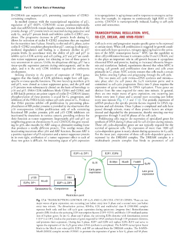
E2F1–3, DP1 E2F7, E2F8
G 1 /S B–Myb MuvB FoxM1 G 2 /M
Early gene
expression Late gene
expression
p130 RB1
MuvB
E2F4, DP1 E2F1–3, DP1
Quiescence G 0 Competence
Restriction point
Fig. 17.4 TRANSCRIPTION CONTROL OF G 1 /S AND G 2 /M CELL CYCLE GENES. There are two
major waves of gene expression, one occurring just before entry into S phase and a second wave just before
entry into mitosis. The dimerization partner, RB-like, E2F, and multivulval class B (DREAM) complex
represses both early and late cell cycle gene expression during quiescence (magenta line). Retinoblastoma
protein (RB) binds to the activating E2F transcription factors (E2F1–E2F3) and blocks E2F-mediated activa-
tion of S-phase genes. In late G 1 phase and S phase, the activating E2Fs dimerize with dimerization partner
1 (DP1) or DP2, bind to the promoters of genes required for DNA synthesis through E2F promoter elements,
and promote their expression. During late S phase, E2F7 and E2F8 will replace E2F1–E2F3 and serve to
repress the expression of the G 1 /S genes when DNA synthesis is completed. The B-MYB transcription factor
binds to the MuvB core when p130, E2F4, and DP are released from the DREAM complex. The B-MYB–
MuvB (MMB) complex recruits FOXM1 to promote the expression of genes in late G 2 phase and M phase.