
Page 226 - Hematology_ Basic Principles and Practice ( PDFDrive )
P. 226
178 Part II Cellular Basis of Hematology
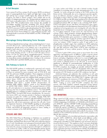
B-Cell Receptor are many cyclins and CDKs, but only a limited number directly
contribute to promoting cell cycle entry and progression (Fig. 17.3).
Every normal B cell has a unique B-cell receptor (BCR) consisting of Typically, the cyclin component binding to the CDK activates the
pairs of immunoglobulin heavy (IgH) and light (IgL) chains. Each kinase activity. Additionally, the cyclin component provides specific-
IgH and IgL has a unique variable region that allows the BCR to ity to the substrate. During G 1 phase, cyclin D (cyclin D1, D2, or
recognize and bind to diverse antigens, both soluble and on the D3) binds to either CDK4 or CDK6. The principal targets of cyclin
surface of antigen-presenting cells. Antigen-induced aggregation of D–CDK4/6 are RB and the RB-related protein p130, which become
7
BCR leads to phosphorylation of ITAMs by the Src family tyrosine monophosphorylated by cyclin D–CDK4/6. Unphosphorylated and
kinase LYN. This phosphorylation event initiates the assembly of monophosphorylated RB can bind to the activating E2F (E2F1–
intracellular signaling molecules, including SYK, PLCγ2, Bruton E2F3) transcription factors and block E2F-dependent gene expres-
tyrosine kinase (BTK), VAV, and the adaptor B-cell linker (BLNK). sion. In accordance with the important functions of cyclin D–CDK4/6
The BCR coreceptor CD19 is also phosphorylated by LYN, leading complexes in leaving quiescence and entering the cell cycle, they are
3
to the recruitment of PI3K, BTK, and AKT. Together, this signaling frequently found overexpressed or mutated in some cancers. At the
2+
leads to the release of intracellular Ca and production of DAG, and G 1- to S-phase transition, E-type cyclins (E1 and E2) bind to and
ultimately leads to activation of RAS-MAPK and PKC–NFκB signal- activate CDK2, which promotes multiple phosphorylation (hyper-
ing pathways. phosphorylation) of RB and dissociation of RB from E2F. Conse-
quently, E2F-dependent gene expression is enabled, leading to the
expression of genes that encode for proteins required for DNA rep-
Macrophage Colony-Stimulating Factor Receptor lication. Cyclin A binds to CDK2 at the end of S phase and promotes
entry into mitosis. CDK2 bound to cyclin E and cyclin A can
The known ligands of macrophage colony-stimulating factor 1 recep- phosphorylate multiple targets that contribute to DNA replication
4
tor (CSF-1R) are CSF-1 and interleukin (IL)-34. CSF-1 was the first and cell cycle progression during S and G 2 phases. Finally, cyclin B
hemopoietic growth factor to be isolated, and it can promote the (B1 and B2) associates with CDK1 (CDC2) and contributes to
growth of pure colonies of macrophages from bone marrow progeni- phosphorylation of many cellular proteins, driving cells through
tors. CSF-1R is an RTK that belongs to the platelet-derived growth mitosis.
factor family. Binding of CSF-1 or IL-34 to CSF-1R leads to dimer- Control of cyclin-CDK activity occurs at many levels. First is the
ization of CSF-1R, which allows autophosphorylation. Numerous appearance and disappearance of different cyclins at specific phases
proteins, such as GRB2, SOS, SFK, CBL, and p85, are recruited to of the cell cycle, which dictates the cyclin–CDK complexes that can
the intracellular domain phosphotyrosines, leading to the activation form in each phase. Regulation at this level is a result of highly regu-
of the PI3K-AKT and RAS-MAPK pathways, among others. lated synthesis and degradation of cyclin messenger RNA (mRNA)
and protein at different points in the cell cycle. A second level of
regulation is afforded by posttranslational modification of CDK
RAS Pathway to Cyclin D kinases, which is often necessary to activate their function. Cyclin
B–CDK1 complexes, for example, are initially inhibited by WEE1
The RAS-MAPK pathway is evolutionarily conserved and controls kinase and are activated by CDC25C phosphatase when cells enter
many fundamental processes, including cell proliferation. RAS mitosis. A third level of regulation is provided by proteins that inhibit
GTPases are activated by many receptors, such as TCR, BCR, and the activity of CDK kinases or cyclin-CDK complexes (see later).
CSF-1R, and recruit MAPK complexes. These complexes are formed Additional kinases and substrates that contribute to cell cycle
by scaffolds, such as KSR (kinase suppressor of RAS), binding to progression include DDK (Dbf4-dependent kinase), PLK (polo-like
the three-tier MAPK module that comprises RAF, mitogen-activated kinase), and Aurora kinases. The DDK CDC7, together with cyclin
protein kinase (MEK), and extracellular signal–regulated kinase E–CDK2, coordinates the initiation of DNA replication. The polo-
(ERK). When activated by RAS (H-RAS, K-RAS, N-RAS), the like kinase PLK1 activates, among others, CDC25C and deactivates
MAPKKK RAF (RAF-1, A-RAF, B-RAF) phosphorylates the WEE1, leading to active cyclin B–CDK1 complexes that drive
MAPKK MEK (MEK1, MEK2), which in turn phosphorylates mitosis, and PLK4 regulates centriole biogenesis during mitosis.
the MAPK ERK (ERK1, ERK2). The principal function of RAS Aurora kinases coordinate mitotic progression through phosphoryla-
in cell cycle induction is to inactivate retinoblastoma protein (RB), tion of multiple proteins that function in chromosome segregation
5
relieving cells from its growth-inhibitory actions. The RAS-MAPK and cytokinesis.
signaling pathway is required to induce complexes of cyclin D1 and
CDK4/CDK6. Activated ERK kinases phosphorylate the transcrip-
tion factors MYC and activator protein 1, leading to transcription CDK INHIBITORS
activation of cyclin D1, CDK4, and CDK6. Notably, PI3K-AKT
and PKC–NFκB pathways cooperate with RAS-MAPK in activating Sharp control of CDK activity is achieved by two major classes of
cyclin D1. Monophosphorylation of RB by cyclin D–CDK4/6 is small polypeptide CDK inhibitors: the INK4 family (inhibitors of
required for cells to leave quiescence and enter the cell cycle. Given CDK4) comprising p16 (INK4A), p15 (INK4B), p18 (INK4C), and
the important functions of RAS-MAPK signaling in cell prolifera- p19 (INK4D); and the CIP/KIP (CDK-interacting protein/kinase
tion, it is not surprising that cancer hijacks this pathway: K-RAS, inhibitor protein) family that comprises p21 (CDKN1A), p27
8
N-RAS, and B-RAF are frequently mutated in cancer, leading to (CDKN1B), and p57 (CDKN1C). All members of the INK4 family
constant activation of the RAS-MAPK pathway. specifically bind to CDK4 and CDK6, inhibiting their kinase activity
by competing with their association with cyclin D. In contrast, CDK
inhibitors of the CIP/KIP family bind to cyclin–CDK complexes and
CYCLINS AND CDKS disturb their activities.
The first inhibitor to be identified and cloned in mammalian cells
The 2001 Nobel Prize in Physiology or Medicine was awarded for was p21 (CIP1, CDKN1A), which binds several different cyclin–
discoveries concerning the control of the cell cycle: Leland Hartwell, CDK complexes. The proteins p27 (Kip1) and p57 (Kip2) were
Tim Hunt, and Paul Nurse discovered CDKs and cyclins that regulate subsequently identified as CDK inhibitors with structural and func-
the cell cycle. Identification and subsequent functional analysis of the tional similarities to p21. The regulation of p21 expression sheds light
9
factors and cofactors involved in mammalian cell cycle regulation on its function : Expression is transcriptionally induced by p53, the
have led to the current view that progression through the cell division tumor suppressor protein activated by DNA damage, and induction
cycle is driven by CDKs. CDKs are serine/threonine protein kinases of p21 expression provides a mechanism for halting cell proliferation
6
that play an essential role in promoting the cell division cycle. There after DNA damage to allow time for damage assessment and repair.