
Page 225 - Hematology_ Basic Principles and Practice ( PDFDrive )
P. 225
Chapter 17 Control of Cell Division 177
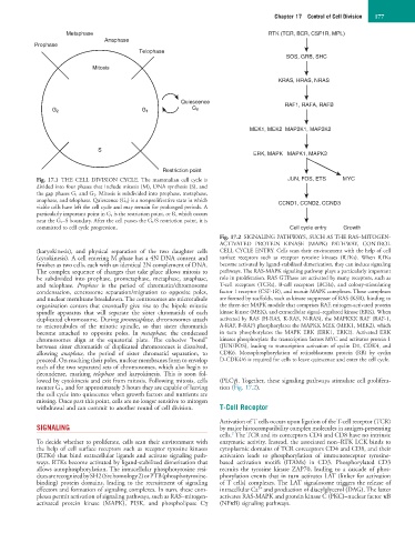
Metaphase RTK (TCR, BCR, CSF1R, MPL)
Anaphase
Prophase
Telophase
SOS, GRB, SHC
Mitosis
KRAS, HRAS, NRAS
Quiescence RAF1, RAFA, RAFB
G 0
G 2 G 1
MEK1, MEK2 MAP2K1, MAP2K2
S
ERK, MAPK MAPK1, MAPK3
Restriction point
Fig. 17.1 THE CELL DIVISION CYCLE. The mammalian cell cycle is JUN, FOS, ETS MYC
divided into four phases that include mitosis (M), DNA synthesis (S), and
the gap phases G 1 and G 2. Mitosis is subdivided into prophase, metaphase,
anaphase, and telophase. Quiescence (G 0) is a nonproliferative state in which CCND1, CCND2, CCND3
viable cells have left the cell cycle and may remain for prolonged periods. A
particularly important point in G 1 is the restriction point, or R, which occurs
near the G 1–S boundary. After the cell passes the G 1/S restriction point, it is
committed to cell cycle progression. Cell cycle entry Growth
Fig. 17.2 SIGNALING PATHWAYS, SUCH AS THE RAS–MITOGEN-
ACTIVATED PROTEIN KINASE (MAPK) PATHWAY, CONTROL
(karyokinesis), and physical separation of the two daughter cells CELL CYCLE ENTRY. Cells scan their environment with the help of cell
(cytokinesis). A cell entering M phase has a 4N DNA content and surface receptors such as receptor tyrosine kinases (RTKs). When RTKs
finishes as two cells, each with an identical 2N complement of DNA. become activated by ligand-stabilized dimerization, they can induce signaling
The complex sequence of changes that take place allows mitosis to pathways. The RAS-MAPK signaling pathway plays a particularly important
be subdivided into prophase, prometaphase, metaphase, anaphase, role in proliferation. RAS GTPases are activated by many receptors, such as
and telophase. Prophase is the period of chromatin/chromosome T-cell receptors (TCRs), B-cell receptors (BCRs), and colony-stimulating
condensation, centrosome separation/migration to opposite poles, factor 1 receptor (CSF-1R), and recruit MAPK complexes. These complexes
and nuclear membrane breakdown. The centrosomes are microtubule are formed by scaffolds, such as kinase suppressor of RAS (KSR), binding to
organization centers that eventually give rise to the bipole mitotic the three-tier MAPK module that comprises RAF, mitogen-activated protein
spindle apparatus that will separate the sister chromatids of each kinase kinase (MEK), and extracellular signal–regulated kinase (ERK). When
duplicated chromosome. During prometaphase, chromosomes attach activated by RAS (H-RAS, K-RAS, N-RAS), the MAPKKK RAF (RAF-1,
to microtubules of the mitotic spindle, so that sister chromatids A-RAF, B-RAF) phosphorylates the MAPKK MEK (MEK1, MEK2), which
become attached to opposite poles. In metaphase, the condensed in turn phosphorylates the MAPK ERK (ERK1, ERK2). Activated ERK
chromosomes align at the equatorial plate. The cohesive “bond” kinases phosphorylate the transcription factors MYC and activator protein 1
between sister chromatids of duplicated chromosomes is dissolved, (JUN/FOS), leading to transcription activation of cyclin D1, CDK4, and
allowing anaphase, the period of sister chromatid separation, to CDK6. Monophosphorylation of retinoblastoma protein (RB) by cyclin
proceed. On reaching their poles, nuclear membranes form to envelop D-CDK4/6 is required for cells to leave quiescence and enter the cell cycle.
each of the two separated sets of chromosomes, which also begin to
decondense, marking telophase and karyokinesis. This is soon fol-
lowed by cytokinesis and exit from mitosis. Following mitosis, cells (PLCγ). Together, these signaling pathways stimulate cell prolifera-
reenter G 1 , and for approximately 3 hours they are capable of leaving tion (Fig. 17.2).
the cell cycle into quiescence when growth factors and nutrients are
missing. Once past this point, cells are no longer sensitive to mitogen
withdrawal and can commit to another round of cell division. T-Cell Receptor
Activation of T cells occurs upon ligation of the T-cell receptor (TCR)
SIGNALING by major histocompatibility complex molecules in antigen-presenting
2
cells. The TCR and its coreceptors CD4 and CD8 have no intrinsic
To decide whether to proliferate, cells scan their environment with enzymatic activity. Instead, the associated non–RTK LCK binds to
the help of cell surface receptors such as receptor tyrosine kinases cytoplasmic domains of TCR coreceptors CD4 and CD8, and their
(RTKs) that bind extracellular ligands and activate signaling path- activation leads to phosphorylation of immunoreceptor tyrosine-
ways. RTKs become activated by ligand-stabilized dimerization that based activation motifs (ITAMs) in CD3. Phosphorylated CD3
allows autophosphorylation. The intracellular phosphotyrosine resi- recruits the tyrosine kinase ZAP70, leading to a cascade of phos-
dues are recognized by SH2 (Src homology 2) or PTB (phosphotyrosine- phorylation events that in turn activates LAT (linker for activation
binding) protein domains, leading to the recruitment of signaling of T cells) complexes. The LAT signalosome triggers the release of
2+
effectors and formation of signaling complexes. In turn, these com- intracellular Ca and production of diacylglycerol (DAG). The latter
plexes permit activation of signaling pathways, such as RAS–mitogen- activates RAS-MAPK and protein kinase C (PKC)–nuclear factor κB
activated protein kinase (MAPK), PI3K, and phospholipase Cγ (NFκB) signaling pathways.