
Page 2293 - Hematology_ Basic Principles and Practice ( PDFDrive )
P. 2293
Chapter 137 Rare Coagulation Factor Deficiencies 2035
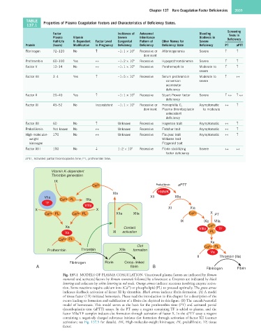
TABLE Properties of Plasma Coagulation Factors and Characteristics of Deficiency States.
137.1
Screening
Factor Incidence of Autosomal Bleeding Tests in
Plasma Vitamin Severe Inheritance Diathesis in Deficiency
Half-Life K–Dependent Factor Level Congenital Pattern of Other Names for Severe
Protein (hours) Modification in Pregnancy Deficiency Deficiency Deficiency State Deficiency PT aPTT
Fibrinogen 72–120 No ↑ ~1 : 1 × 10 6 Recessive or Afibrinogenemia Severe ↑ ↑
dominant
Prothrombin 60–100 Yes ↔ ~1 : 2 × 10 6 Recessive Hypoprothrombinemia Severe ↑ ↑
Factor V 12–14 No ↔ ~1 : 1 × 10 6 Recessive Parahemophilia Moderate to ↑ ↑
severe
Factor VII 3–4 Yes ↑ ~1 : 5 × 10 5 Recessive Serum prothrombin Moderate to ↑ ↔
conversion severe
accelerator
deficiency
Factor X 20–40 Yes ↑ ~1 : 1 × 10 6 Recessive Stuart-Prower factor Severe ↑ ↔ ↑ ↔
deficiency
Factor XI 45–52 No Inconsistent ~1 : 1 × 10 6 Recessive or Hemophilia C, Asymptomatic ↔ ↑
dominant Plasma thromboplastin to moderate
antecedent
deficiency
Factor XII 60 No ↑ Unknown Recessive Hageman trait Asymptomatic ↔ ↑
Prekallikrein Not known No ↔ Unknown Recessive Fletcher trait Asymptomatic ↔ ↑
High-molecular- 170 No ↔ Unknown Recessive Flaujeac trait Asymptomatic ↔ ↑
weight Williams trait
kininogen Fitzgerald trait
Factor XIII 150 No ↓ 1 : 2 × 10 6 Recessive Fibrin stabilizing Severe ↔ ↔
factor deficiency
aPTT, Activated partial thromboplastin time; PT, prothrombin time.
Vitamin K−dependent
Thrombin generation
IX
Ca 2+ Prekallikrein aPTT
HMWK
XIa
VIIa XII XIIa
Ca 2+ PL IXa
TF
VIIIa
X X XI XIa
Ca 2+ PL Ca 2+ PL XIIa XIIa Ca 2+ X PT
IX IXa VIIa
Xa Contact VIIIa Ca 2+ TF
Va XI activation PL
Ca 2+ PL
Xa
Clot Ca 2+ PL
Prothrombin Thrombin XIIa formation Va
II Thrombin (IIa)
Fibrinogen Fibrin Cross−linked
A fibrin B
Fibrinogen Fibrin
Fig. 137.1 MODELS OF PLASMA COAGULATION. Unactivated plasma factors are indicated by Roman
numerals and activated factors by Roman numerals followed by a lowercase a. Enzymes are indicated by black
lettering and cofactors by white lettering in red ovals. Orange arrows indicate reactions involving enzyme activa-
2+
tion. Some reactions require calcium ions (Ca ) or phospholipid (PL) to proceed optimally. The green arrow
indicates feedback activation of factor XI by thrombin. Black arrows indicate fibrin formation. (A) A model
of tissue factor (TF) initiated hemostasis. Please read the introduction to this chapter for a description of the
events leading to formation and stabilization of a fibrin clot depicted in this figure. (B) The cascade/waterfall
model of hemostasis. This model serves as the basis for the prothrombin time (PT) and activated partial
thromboplastin time (aPTT) assays. In the PT assay a reagent containing TF is added to plasma, and the
factor VIIa/TF complex induces clot formation through activation of factor X. In the aPTT assay a reagent
containing a negatively charged substance initiates clot formation through activation of factor XII (contact
activation; see Fig. 137.5 for details). HK, High-molecular-weight kininogen; PK, prekallikrein; TF, tissue
factor.