
Page 2299 - Hematology_ Basic Principles and Practice ( PDFDrive )
P. 2299
Chapter 137 Rare Coagulation Factor Deficiencies 2041
relatively low (<2%). It is reasonable to maintain the factor VII, IX, Factor V
and X levels at less than 150% of normal to reduce risk. Dental Heavy chain B domain Light chain
procedures or minor hemorrhage may respond to antifibrinolytic
1
therapy with ε-amino caproic acid. Prothrombin deficiency may also R709
be treated with FFP for bleeding episodes or surgical intervention (15
to 20 mL/kg loading dose followed by 3 mL/kg/day). Because of the Heavy chain Light chain
long half-life, additional doses may not be required in all situations. R1018
Cryoprecipitate is not a source of prothrombin, and plasma pro-
thrombin levels do not increase after infusion or inhalation of des- Heavy chain Light chain
mopressin (1-desamino-8-D-arginine vasopressin [DDAVP]).
The postviral hypoprothrombinemia seen in young children often R1545
spontaneously resolves. Intravenous immunoglobulin has been effec- Factor Va
tive is this population. Treatment of acquired prothrombin deficiency Heavy chain Ca 2 Light chain
associated with lupus anticoagulants in patients with autoimmune Factor V-short
diseases often requires immune suppression. Steroids are effective in
most patients, although many relapse during weaning or after stop- Heavy chain Light chain
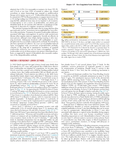
ping treatment. Subsequent treatment with azathioprine or cyclo- Fig. 137.4 SCHEMATIC DIAGRAMS OF HUMAN FACTOR V AND
phosphamide has successfully eradicated the antibody. Rituximab has FACTOR Va. In factor V the heavy and light chains (yellow) are separated
also been reported to be effective. In a rare case of quinidine-induced by a central B domain (white, amino acids 710 to 1545) that contains a basic
lupus anticoagulant with concomitant antiprothrombin antibody, region (blue, amino acids 963 to 1008) and acidic region (red, amino acids
cessation of the drug led to spontaneous resolution of acquired 1493 to 1537) Removal of the B domain by thrombin to generate factor Va
prothrombin deficiency, but not the lupus anticoagulant. The low involves sequential cleavages after Arginine 709, 1018 and 1545. Factor
prothrombin activity in these patients may protect them from throm- V-short is the product of an alternatively spliced factor V mRNA that lacks
bosis, as suggested by reports of thrombosis after successful eradica- residues 756 through 1458, which comprise most of the B domain including
tion of the antiprothrombin antibody. the basic region. In factor V-short, the loss of the B domain basic region leaves
the acidic region free to bind to TFPI.
FACTOR V DEFICIENCY (OMIM 227400)
In 1943 Quick reported that aged plasma clotted more slowly than low platelet factor V and normal plasma factor V levels. In this
fresh plasma in a PT assay, and proposed that a labile factor distinct syndrome, excessive proteolysis of α-granule proteins is caused
from prothrombin was required for coagulation. At the same time, by overexpression of urokinase. Platelet factor V activity is also
Owren noted that a patient with a lifelong bleeding problem lacked reduced in factor V New York, but the underlying mechanism is
a plasma factor that, unlike prothrombin, did not adsorb onto alu- not known.
minum hydroxide. Owren’s patient was deficient in the labile factor The autosomal dominant condition East Texas bleeding disorder
described by Quick, which is now called factor V. Moderate to severe is caused by an A2440G nucleotide substitution in exon 13 of the
congenital factor V deficiency (1%–10% of normal level) occurs in factor V gene. The substitution increases levels of the alternatively
1 in 1 million persons (Table 137.1). 13,14 Full length factor V, a spliced mRNA encoding Factor V-short (Fig. 137.4), leading to
homolog of factor VIII, is the 330,000-Da precursor of the cofactor increased plasma levels of this protein. In East Texas bleeding disorder
factor Va, which facilitates prothrombin activation by factor Xa on plasma levels of a key regulator of coagulation, tissue factor pathway
phospholipid surfaces (Fig. 137.1). 13,14 During coagulation, the inhibitor (TFPI), are 10-fold higher than normal. TFPI-mediated
B-domain of factor V is removed by thrombin or factor Xa to produce inhibition of factor Xa and the factor VIIa-tissue factor complex likely
factor Va (Fig. 137.4). An approximately 250,000-Da form of factor contributes to the bleeding. In factor V, the B-domain contains basic
13
V lacking 703 amino acids from the B-domain is a minor constituent and acid regions that are thought to bind to each other (Fig. 137.4).
in normal plasma (Fig. 137.4). Referred to as factor V-short (or factor In factor V-short, the basic region is missing, leaving the acidic region
V East Texas), it is a product of an alternatively spliced mRNA. Most free to bind to TFPI, stabilizing it in plasma. A similar bleeding
(80%) factor V in blood is in plasma, with the remainder stored in disorder was observed in a Dutch family with a C2588G substitution
13
platelet α-granules. In humans, platelet factor V is primarily of in exon 13 of the factor V gene, leading to alternative mRNA splicing
plasma origin. It is taken up by megakaryocytes in a process requiring and loss of 632 amino acids from the B domain (factor V Amster-
low-density lipoprotein receptor-related protein-1 (LRP-1), then dam). These patients also have markedly elevated plasma TFPI levels.
converted to a partially activated form. 13 Acquired factor V deficiency may occur with liver disease, DIC,
Severe factor V deficiency is an autosomal recessive trait with myleoproliferative disorders or systemic amyloidosis. Patients with
undetectable (<1% of normal) plasma factor V. In moderate defi- acquired antibodies to factor V may have bleeding that can be severe,
ciency the level is between 1% and 10% of normal (Table 137.2). 13,14 or may be asymptomatic. Alloantibodies to factor V were often
More than 100 factor V gene mutations have been described in factor associated with exposure to topical bovine thrombin during surgery,
13
V–deficient patients. Nonsense and frameshift mutations and splice a problem that rarely occurs now because recombinant human
variants are common and are distributed throughout the gene. Mis- thrombin preparations are used. Alloantibodies to factor V also occur
sense mutations cluster in the A2 and C2 domains, 13,14 and usually in some factor V–deficient patients exposed to human plasma. Factor
result in abnormal polypeptides that are degraded within the cell, V autoantibodies may form after surgery or blood transfusions or
resulting in low plasma factor V antigen (CRM − deficiency). While with cancer, autoimmune disorders, or therapy with β-lactam or
it is estimated that approximately 25% of cases of factor V deficiency aminoglycoside antibiotics.
are CRM+ mutations (plasma antigen exceeds activity), only two There is striking variability in bleeding symptoms among patients
have been characterized. The Ala221Val (factor V New Brunswick) with severe factor V deficiency. 13,14 Significant bleeding does occur,
and His147Arg substitutions appear to reduce factor Va activity by but the frequency tends to be less than in patients lacking factor VIII
affecting protein stability. or factor IX. Several factors may contribute to the variability. Some
Combined deficiency of factor V and factor VIII is caused by patients with mild bleeding symptoms despite low plasma factor V
mutations in proteins required for secretion of both (see Combined levels have sufficient platelet factor V to support thrombin genera-
Factor V and Factor VIII Deficiency). 13,14 A few patients have been tion. This implies that their factor V is unstable in plasma, but can
13
described with abnormalities specific to platelet factor V. The be taken up by platelets. Patients with severe bleeding may lack
Quebec platelet disorder is an autosomal dominant condition with plasma and platelet factor V. In one such individual treated with