
Page 2336 - Hematology_ Basic Principles and Practice ( PDFDrive )
P. 2336
2078 Part XII Hemostasis and Thrombosis
Protein C Pathway TABLE
140.4 Types of Inherited Protein C Deficiency
Type Antigen Activity
Thrombin generation
COAGULATION inhibited I Low Low
II Normal Low
Thrombin
generation
II A Va Vi APC has a half-life in the circulation of about 15 minutes, whereas
Thrombin Protein C P Va L Vi thrombin has a half-life of about 10 seconds. APC is inhibited by
inactivation activation C protein C inhibitor and α 1-proteinase inhibitor (α 1-antitrypsin),
PS both of which are relatively slow inhibitors. Only the activity of
IIa protein C inhibitor is enhanced by heparin, but both inhibitors
PC
TM EPCR appear to contribute to APC inhibition in vivo.
Protein C deficiency can be inherited or acquired. Like antithrom-
bin deficiency, protein C deficiency is inherited in an autosomal-
dominant fashion and has a proven association with venous
5
thrombosis. Based on studies in healthy blood donors, heterozygous
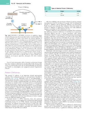
Fig. 140.2 PROTEIN C PATHWAY. Activation of coagulation triggers protein C deficiency can be found in 1 in 200 to 500 of the adult
thrombin (IIa) generation. Excess thrombin binds to thrombomodulin (TM) population, but many of these individuals do not have a history of
on the endothelial cell surface. Once bound, the substrate specificity of thrombosis. Thus the phenotypic expression of hereditary protein C
thrombin is altered so that it no longer acts as a procoagulant but becomes deficiency is highly variable and may depend on other, as yet unrec-
a potent activator of protein C (PC). Endothelial protein C receptor (EPCR) ognized, modifying factors. In contrast to antithrombin deficiency in
binds PC and presents it to thrombomodulin-bound thrombin, where it is which the homozygous state is embryonic lethal, homozygous or
activated. Activated protein C (APC), together with its cofactor, protein S doubly heterozygous protein C deficiency can occur. The prevalence
(PS), binds to the activated platelet-surface and proteolytically degrades factor of homozygous protein C deficiency is estimated to be 1 in 160,000
Va (Va) into inactive fragments (Vi). Because factor Va is a critical component to 360,000 births. Newborns with these disorders may present with
of the prothrombinase complex, factor Va inactivation by APC attenuates purpura fulminans characterized by widespread thrombosis.
thrombin generation. Because factor Va Leiden (FVa L ) is resistant to inactivation Individuals with heterozygous protein C deficiency can develop
by APC, patients with the factor V Leiden mutation have reduced capacity to skin necrosis upon initiation of treatment with vitamin K antagonists,
regulate thrombin generation. (From Anderson JA, Weitz JI: Hypercoagulability such as warfarin. Typically, skin lesions are found on the extremities,
and uncommon vascular diseases. In Jaff MR, White CJ, editors: Vascular disease: breasts, or trunk. Starting as erythematous macules, the central
Diagnostic and therapeutic approaches, Minneapolis, MN, 2011, Cardiotext regions of the cutaneous lesions become purpuric and then necrotic
Publishing.)
over a period of hours unless protein C is administered. Biopsies
reveal fibrin thrombi within the vessels of the skin associated with
interstitial hemorrhage. The skin lesions are clinically and histologi-
Users of oral contraceptive pills or hormone replacement therapy cally similar to those seen in infants with purpura fulminans and are
may have moderate reductions in antithrombin levels; during preg- attributable to the transient hypercoagulable state that is induced by
nancy, the antithrombin levels do not fall significantly but decreases warfarin, thereby explaining why they occur early during the course
are found in preeclampsia and pregnancy-induced hypertensive of warfarin therapy. The half-life of protein C is short and similar to
illnesses. that of factor VII. Starting warfarin in patients with protein C defi-
ciency causes a further reduction in protein C levels, particularly if
loading doses of warfarin are given. Thus the activity of the natural
Protein C Deficiency anticoagulant protein C pathway is compromised before warfarin
lowers vitamin K–dependent procoagulant proteins, particularly
The protein C pathway is an important natural anticoagulant prothrombin and factor X, into the range required for its antithrom-
pathway. This on-demand pathway is activated when thrombin is botic effects. Warfarin-induced skin necrosis has also been reported
generated (Fig. 140.2). Thrombin binds to thrombomodulin, a in association with acquired deficiency of protein C.
transmembrane thrombin receptor found on the surface of endothe- Hereditary protein C deficiency can be further delineated into two
lial cells. Once bound to thrombomodulin, the substrate specificity subtypes using immunologic and functional assays (Table 140.4).
of thrombin is altered so that it no longer serves as a procoagulant Most functional assays use Protac, a protease isolated from the venom
enzyme, but becomes a potent activator of protein C, a vitamin K– of the copperhead snake, to activate protein C in plasma. The enzy-
dependent glycoprotein. Thus thrombin bound to thrombomodulin matic activity of APC can then be assayed directly using an APC-
activates protein C 1000-fold more efficiently than free thrombin. directed synthetic substrate, or it can be indirectly quantified by
The endothelial cell protein C receptor (EPCR), another transmem- measuring the extent of prolongation of the activated partial throm-
brane receptor on the endothelial cell surface, binds protein C and boplastin time (aPTT). The most common form of hereditary protein
presents it to the thrombin–thrombomodulin complex for activation, C deficiency is the classic or type I deficiency state. This disorder
thereby producing an additional 20-fold increase in the rate of protein reflects reduced synthesis of a normal protein and is characterized by
C activation. The physiologic importance of thrombomodulin and a parallel reduction in protein C antigen and activity, resulting in a
EPCR is highlighted by the fact that deficiency of either receptor quantitative deficiency due to reduced synthesis or stability of protein
results in embryonic lethality in mice. C. A variety of genetic defects can produce type I protein C deficiency,
Activated protein C (APC) dissociates from this activation including promoter mutations, splice-site abnormalities, in-frame
complex and acts as an anticoagulant by proteolytically degrading and deletions, frameshift deletions, in-frame insertions, and frameshift
inactivating factors Va and VIIIa, thereby attenuating thrombin insertions in the protein C gene (PROC), but missense or nonsense
generation. For efficient inactivation of factors Va and VIIIa, APC mutations are the most common.
must bind to protein S, its cofactor. This interaction facilitates APC Type II protein C deficiency reflects synthesis of a dysfunctional
binding to activated cell surfaces, particularly the platelet surface, protein and is characterized by normal protein C antigen with
where factors Va and VIIIa are localized. reduced functional activity, a qualitative deficiency. Most type II