
Page 236 - Hematology_ Basic Principles and Practice ( PDFDrive )
P. 236
188 Part II Cellular Basis of Hematology
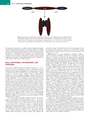
Prodomain Large Small
Asp-X Asp-X
Fig. 18.4 MATURE CASPASES ARE FORMED BY PROTEOLYTIC PROCESSING OF PROCASPASES
TO DIVIDE LARGE AND SMALL SUBUNITS AND REMOVE N-TERMINAL PEPTIDES. Caspase
substrate motifs at cleavage sites enable sequential caspase activation, or in the case of initiator caspases,
autoactivation. Caspase dimers are assembled from two large and two small subunits. Asp, Aspartate.
for processing. Interactions at a dimer interface (induced proximity proteolytic activity. Eventual dissociation from the apoptosome due
or induced conformation model) reorient and stabilize the binding to cleavage of the prodomain results in loss of proteolytic activity.
pocket conformation of these caspases. Normally monomeric, these Both pathways converge with proteolytic activation of caspase-3 by
zymogens are distinguished by the presence of a long prodomain caspase-8, -9, or -10.
that serves as a docking site for recruitment into a self-activating Inflammasomes are large multiprotein complexes, visible as
complex. Protein associations within these complexes are built around speck structures in stimulated cells, that are dedicated to activating
homomeric interactions between three binding cassettes, death (DD), caspase-1 in response to molecular cues from infectious pathogens
death effector (DED), and CARD domains. (pathogen-associated molecular patterns [PAMPs]) or endogenous
5
signals (danger-associated molecular patterns [DAMPs]). A family
DISCS, APOPTOSOMES, INFLAMMASOMES, AND of nucleotide-binding oligomerization domain (Nod)-like receptors
(NLRs) detect PAMPs and DAMPs through leucine-rich repeats
PIDDOSOMES (LRRs), triggering oligomerization of Nod domains. A third,
N-terminal, region contains one of several protein interaction motifs,
Four distinct caspase-activating assemblies are known (Fig. 18.5). including CARD and pyrin domains, for direct interaction with
Caspase-8 and -10 are engaged by a family of cell surface receptors caspase-1 or via an adaptor protein, apoptosis-associated speck-like
known as death receptors, including tumor necrosis factor receptor protein containing a CARD (ASC) (see Fig. 18.5C). The three
3,4
1 (TNFR1), Fas/CD95, TNFRSF10A, and TNFRSF10B. Ligand NLR inflammasomes characterized to date are named after the NLR
binding to trimerized death receptors induces conformational protein involved. NLRP1 and NLRC4 recognize bacterial muramyl
changes that promote binding of adaptor proteins, Fas-associated dipeptide and flagellins, respectively, while NLRP3 recognizes mul-
death domain protein (FADD) and TNFR1-associated death domain tiple stimuli, including saturated fatty acids, bacterial RNA, and
protein (TRADD), to the cytoplasmic tail of the death receptor urate crystals. Non-NLR family inflammasome proteins, AIM2 and
by dimerization of homologous death domains (DDs) from each PYRIN, recognize cytosolic dsDNA and modified Rho GTPases,
molecule. A second interaction domain in FADD, a DED, binds to respectively. The inflammasome scaffold is postulated to trigger
a similar DED in the prodomain of caspase-8/10, leading to caspase caspase-1 activity according to the induced proximity model. An
dimerization and localized autocatalysis. The prodomain of caspase- inflammatory cell death termed pyroptosis is initiated by inflamma-
8/10 is severed during processing, dispersing active caspases to cellular some activation. Inflammasome activation may also be triggered by
substrates. Analysis of unprocessed caspase-8 dimers demonstrated viral PAMPs. RNA viruses are recognized by retinoic acid-inducible
that active sites can be formed in the absence of processing, stabilized gene-1 (RIG-I) and RIG-I-like-receptor (RLR) helicases, which
by hydrophobic interactions at the dimer interface. The death recep- oligomerize upon binding viral RNA and translocate to the interface
tor, FADD, and caspase complex is known as the death-inducing between mitochondrial membranes and mitochondria-associated
signaling complex (DISC) (see Fig. 18.5A). In the case of TNFR1, membranes. Binding to the mitochondrial antiviral signaling protein
additional complexes are involved in nuclear factor kappa-B (NFκB) (MAVS), a tail-anchored membrane adaptor protein, activates IRF3
signaling and necroptosis. and NFκB transcription of antiviral responses. 6
The second caspase-activating assembly platform, the apopto- Caspase-2 is activated following genotoxic damage via a p53-
some, is specialized for activating caspase-9 and -7, which have inducible protein, p53-induced protein with a death domain (PIDD),
CARD-type prodomains. Formation of the cytoplasmic apoptosome in a complex known as the PIDDosome. Similar to death receptors,
is initiated by release of the soluble electron carrier, cytochrome death domains in PIDD bind to an adaptor protein, RAIDD, which
c, from mitochondria. Cytochrome c binds to an adaptor protein, in turn recruits caspase-2 through death effector domain interactions,
apoptotic protease activating factor 1 (APAF-1), enabling adenosine generating a large (>670 kDa) multiprotein complex (see Fig. 18.5D).
triphosphate (ATP)/dATP-dependent oligomerization of APAF-1 PIDD also activates NFκB downstream of DNA damage responses
in a heptameric wheel and exposure of its own CARD domain through competing interactions with the receptor-interacting protein
(see Fig. 18.5B). Docking of caspase-9 to the apoptosome through 1 (RIP1) serine/threonine kinase and I-kappa-B kinases (IKK) scaf-
CARD–CARD interactions is both necessary and sufficient for fold, NFκB essential modulator (NEMO).