
Page 2517 - Hematology_ Basic Principles and Practice ( PDFDrive )
P. 2517
2240 Part XIII Consultative Hematology
hypertension and vascular deformities. Over 80% of bleeding epi- Being an acute phase reactant, fibrinogen synthesis is generally
sodes in patients with cirrhosis are a result of variceal bleeding. preserved unless liver disease is severe. Acquired dysfibrinogenemia,
Hepatic dysfunction leads to reduced synthesis of most coagula- however, has been described in approximately 75% of patients with
tion factors. The number and degree of clotting factor deficiencies chronic liver disease, acute liver failure, and cirrhosis but is not
reflect the severity of liver damage. Factor VII levels, having the thought to contribute significantly to bleeding. Aberrant polymeriza-
shortest half-life (6 hours) of the coagulation factors, often decline tion of fibrin monomers may be related to excess sialic acid residues
early and are reflected by prolongation of the prothrombin time (PT)/ on fibrinogen, interfering with the activity of thrombin. Laboratory
international normalized ratio (INR). Conversely, factor VIII and findings of dysfibrinogenemia include elevated PT, PTT, or thrombin
vWF levels may be normal or elevated in liver disease because of time, low or normal fibrinogen by immunologic assay and reduced
upregulated compensatory extrahepatic synthesis or impaired hepatic fibrinogen by functional assay.
clearance. The presence of hyperfibrinolysis in liver disease and its contribu-
Reductions in factors II, VII, IX, and X in patients with liver tion to bleeding risk is controversial. Triggers of increased fibrinolysis
disease may also result from vitamin K deficiency caused by malnutri- may involve release of tissue plasminogen activator (t-PA) in the
tion, malabsorption, use of antibiotics, or biliary tract obstruction. setting of infection or surgery, reabsorption of ascitic fluid with
For these coagulation factors, vitamin K is required as a cofactor in fibrinolytic activity, and altered synthetic or metabolic functions of
γ-carboxylation, a process that converts inactive precursors to biologi- the liver. With the exception of t-PA and plasminogen activator
cally active factors. inhibitor-1 (PAI-1), all fibrinolytic and antifibrinolytic proteins are
Defects in coagulation factors are suggested by prolonged PT/INR synthesized in the liver. Decreased hepatic clearance of t-PA and
and partial thromboplastin time (PTT) measurements and confirmed reduced synthesis of α2 antiplasmin and thrombin-activatable fibri-
by individual factor levels. However, these routine screening tests of nolysis inhibitor favor an increase in circulating plasmin and a
coagulation do not identify patients at risk of bleeding. Both the PT hyperfibrinolytic state in cirrhosis. Available laboratory tests cannot
and INR have been incorporated into prognostic indices (Child-Pugh adequately assess the overall activity of profibrinolytic and antifibri-
and Model of End-stage Liver Disease scores) as markers of synthetic nolytic components. Shortened whole blood euglobulin clot lysis
dysfunction to estimate the severity of liver disease and stratify time and elevated levels of D-dimer, fibrin, and fibrinogen degrada-
patients for transplant, respectively. Factor V levels have been studied tion products are suggestive of increased fibrinolysis. These abnormal
as a prognostic indicator in acute fulminant hepatic failure. Acute laboratory indices have been observed in nonbleeding patients but
liver failure is associated with more pronounced elevations in the are seen more frequently in bleeding patients and have been reported
INR, yet is associated with less spontaneous bleeding than chronic to correlate with GI bleeding, severity of liver failure, and variceal
liver failure reflecting the importance of hemodynamics (portal size. Hyperfibrinolysis may theoretically aggravate bleeding through
hypertension) on the risk of bleeding. consumption of coagulation factors, inhibition of fibrin polymeriza-
Despite their routine use in clinical practice, several significant tion, and reduced platelet aggregation via degradation of vWF and
limitations exist in applying the PT/INR or PTT in the context of glycoprotein Ib and α IIb β 3 . Hyperfibrinolysis likely plays a more
liver disease. First, the INR has not been validated for patents with important role in hemostasis during liver transplantation. Conversely,
cirrhosis. Second, there is no evidence that demonstrates correcting patients with acute hepatic failure show evidence of impaired fibri-
these abnormal values with plasma or procoagulant agents prevents nolysis with elevated PAI-1 levels and decreased plasminogen.
bleeding or improves outcomes. Several reasons may account for the
lack of correlation between PT/INR or PTT with bleeding. Liver
disease results in deficiencies of procoagulant proteins but also defi- TREATMENT OF LIVER DISEASE–RELATED BLEEDING
ciencies in the natural anticoagulant proteins, including antithrombin
and proteins C and S. PT/INR and PTT assays reflect procoagulant Treatment and correction of asymptomatic hemostatic abnormalities
protein levels only and do not reflect alterations in anticoagulant in patients with liver disease is generally not indicated and potentially
proteins, the role of the endothelium and platelet number or func- harmful. Interventions may be indicated when there is active bleeding
tion. Small studies have demonstrated normal thrombin generation or before a planned invasive procedure. Most of the evidence for the
in cirrhotic patients and patients with acute liver failure and pro- prevention of bleeding in patients with chronic liver disease is based
longed PT and PTTs. Tests of thrombin generation (e.g., thrombo- on studies of the perioperative management of patients undergoing
elastography, rotational thromboelastometry) have been studied in liver transplantation.
liver transplantation to assess hemostasis and guide transfusions but RBC transfusions should be provided to maintain an adequate
not to predict the risk of bleeding in patients with liver disease hemoglobin or hematocrit levels and for symptomatic anemia. A
undergoing other invasive procedures. randomized controlled trial demonstrated that a restrictive RBC
transfusion threshold in patients with an acute upper GI bleed reduced
rebleeding and increased survival. In general, platelet transfusions are
not indicated for isolated thrombocytopenia in the absence of bleed-
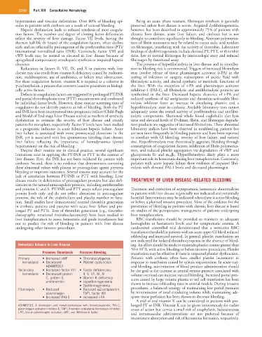
Hemostatic Balance in Liver Disease ing. An effort should be made to maintain platelet counts greater than
9
50 × 10 /L with active bleeding or before invasive procedures. Platelet
Promotes Thrombosis Promotes Bleeding transfusion may be effective if there is suspected platelet dysfunction.
Primary • Increased vWF • Thrombocytopenia Patients with cirrhosis often have smaller platelet increments in
hemostasis • Decreased • Platelet dysfunction response to transfusion caused by splenic sequestration. In acute vari-
ADAMTS13 ceal bleeding, minimization of blood product administration should
Secondary • Increased factor VIII • Factor deficiencies: be the goal as the increase in central venous pressure associated with
hemostasis • Decreased protein II, V, VII, IX, XI volume overload can increase variceal bleeding. Increased portal pres-
C, protein S, • Vitamin K deficiency sures caused by large volume plasma or red cell transfusion has been
antithrombin • Hypofibrinogenemia shown to increase rebleeding rates in animal models. During invasive
• Dysfibrinogenemia
Fibrinolysis • Reduced • Reduced α2-antiplasmin, procedures, a balanced strategy of maintaining low portal pressures
plasminogen TAFI, factor XIII by minimization of total circulating volume while maintaining ade-
• Increased PAI-1 • Increased t-PA quate tissue perfusion has been shown to decrease bleeding.
A trial of oral vitamin K can be considered in patients with pro-
ADAMST13, A disintegrin and metalloproteinase with thrombospondin; PAI-1, longed PT or INR. Vitamin K can be given intravenously for earlier
plasminogen activator inhibitor-1; TAFI, thrombin activatable fibrinolysis inhibitor; onset of action but carries a small risk of anaphylaxis. Subcutaneous
t-PA, tissue plasminogen activator; vWF, von Willebrand factor. and intramuscular administrations are not preferred because of
inconsistent absorption and risk of hematoma formation, respectively.