
Page 2519 - Hematology_ Basic Principles and Practice ( PDFDrive )
P. 2519
2242 Part XIII Consultative Hematology
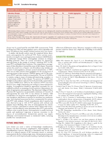
Hemostatic Indices in Liver Disease
Laboratory Changes PT PTT TCT Fib Clauss Plt Platelet Aggregation FVII DD ELT
Thrombocytopenia N N N N N ↓ N N N N
Platelet dysfunction N N N N N N abnormal N N N
Vitamin K deficiency a ↑ ↑ N N N N N ↓ N N
Factor deficiency ↑ ↑ N N N N N ↓ N N
Hypofibrinogenemia N/↑ N/↑ ↑ ↓ ↓ N N N N N
Dysfibrinogenemia N/↑ N/↑ ↑ N ↓ N N N N N
Hyperfibrinolysis N/↑ N/↑ N/↑ N/↓ N/↓ N N ↓ ↑ ↓
DIC N/↑ N/↑ N/↑ ↓ ↓ ↓ N N/↓ ↑ ↓
a Differentiating between vitamin K deficiency and liver disease can be challenging with conventional laboratory tests. If available, performing a factor II assay with and
without Echis venom (factor II biologic and factor II Echis) may be useful. Ecarin is derived from Echis carinatus snake venom and can activate prothrombin irrespective
of γ-carboxylation. Factor II activity (biologic) is reduced in both vitamin K deficiency and liver disease. In contrast, the factor II Echis is reduced in liver disease but is
normal in vitamin K deficiency.
Clauss, Clauss fibrinogen; DD, D-dimer; DIC, disseminated intravascular coagulation; ELT, euglobulin lysis time (measure of fibrinolysis); Fib, fibrinogen; FVII, factor VII
functional assay; N, normal; Plt, platelet; PT, prothrombin time; PTT, partial thromboplastin time; TCT, thrombin clotting time.
disease may be complicated by unreliable INR measurements. Trials refinement of laboratory assays. Moreover, strategies to safely manage
involving non-VKA oral anticoagulants such as direct thrombin and patients with liver disease and a high-risk of bleeding or thrombosis
factor Xa inhibitors have usually excluded patients with liver disease. are required.
A similar risk–benefit analysis must be completed before thera-
peutic anticoagulation for confirmed VTE. Expert opinion recom-
mends screening for varices and appropriate treatment with β-blockers SUGGESTED READINGS
or endoscopic therapy before anticoagulant initiation to mitigate
bleeding potential. There are several indications for therapeutic Afdhal NH, Giannini EG, Tayyab G, et al: Eltrombopag before proce-
anticoagulation in the setting of cirrhosis, including DVT or PE, dures in patients with cirrhosis and thrombocytopenia. N Engl J Med
acute PVT, and Budd-Chiari syndrome. The comprehensive manage- 367:716–724, 2012.
ment of these disorders is beyond the scope of this chapter. Treatment Boyer TD, Habib S: Big spleens and hypersplenism: fix it or forget it? Liver
options for portal vein or hepatic vein thromboses may include one Int 35:1492–1498, 2015.
or more of anticoagulation, systemic or local thrombolysis, or TIPS. Clevenger B, Mallett SV: Transfusion and coagulation management in liver
There are no truly evidence-based guidelines for the management of transplantation. World J Gastroenterol 20:6146–6158, 2014.
anticoagulation in these patients. LMWH appears safe for the treat- Dieterich DT, Spivak JL: Hematologic disorders associated with hepatitis C
ment of PVT and often results in recanalization. The use of LMWHs virus infection and their management. Clin Infect Dis 37:533–541, 2003.
is complex owing to issues of dosing and monitoring, secondary to Ferro D, Celestini A, Violi F: Hyperfibrinolysis in liver disease. Clin Liver
the hemostatic profile of liver disease. The potency of LMWH may Dis 13:21–31, 2009.
be higher in liver disease and anti-Xa levels potentially underestimate Garcia-Tsao G, Sanyal AJ, Grace ND, et al: Prevention and management of
the actual LMWH mass. VKAs for the treatment of PVT have been gastroesophageal varices and variceal hemorrhage in cirrhosis. Hepatology
shown to have a high complication rate owing to a narrow therapeutic 46:922–938, 2007.
window. Anticoagulation should be approached cautiously but not Gonzalez-Casas R, Jones EA, Moreno-Otero R: Spectrum of anemia associ-
withheld needlessly as mounting clinical experience demonstrates the ated with chronic liver disease. World J Gastroenterol 15:4653–4658,
safety of appropriate anticoagulation. The American College of Chest 2009.
Physician 2012 guidelines recommended a minimum of 3 months of Haas T, Fries D, Tanaka K, et al: Usefulness of standard plasma coagulation
anticoagulation for acute PVT in the absence of cirrhosis, with tests in the management of perioperative coagulopathic bleeding: is there
consideration for long-term anticoagulation in patients with persis- any evidence? Br J Anaesth aeu303, 2014.
tent thrombotic risk factors or concerns for extension into mesenteric Kearon C, Akl E, Comerota A: Antithrombotic therapy for VTE disease:
veins. Individualized assessment has been recommended for patients antithrombotic therapy and prevention of thrombosis: ACCP evidence-
with PVT with cirrhosis. Overall recanalization after 6 months has based clinical practice guidelines. Chest 141:e419S–e494S, 2012.
been observed in up to 75% of PVT patients with cirrhosis and Lee WM, Larson AM, Stravitz RT: AASLD position paper: the management
minimal major bleeding. The value of anticoagulation for chronic of acute liver failure: update 2011. Hepatology 55:965–967, 2011.
PVT is uncertain, particularly in patients with cirrhosis with varices. Lisman T, Porte RJ: Hemostatic problems in chronic and acute liver disease.
Hepatic vein thrombosis is more uncommon than PVT, and even less Hemostasis and thrombosis, 2014, pp 271–283.
evidence exists to guide therapy. Anticoagulation is usually instituted Marks PW: Hematologic manifestations of liver disease. Semin Hematol
acutely and maintained long-term to prevent recurrence in the 50:216–221, 2013.
absence of contraindications. Symptomatic patients may require Molenaar IQ, Warnaar N, Groen H, et al: Efficacy and safety of antifibrino-
additional interventional therapies as mentioned earlier, including lytic drugs in liver transplantation: a systematic review and meta-analysis.
possible liver transplantation. Am J Transplant 7:185–194, 2007.
Moore C, Levitsky J: The current state and future prospects of chronic
hepatitis C virus infection treatment. Curr Infect Dis Rep 16:413, 2014.
FUTURE DIRECTIONS Northup PG, Caldwell SH: Coagulation in liver disease: a guide for the
clinician. Clin Gastroenterol Hepatol 11:1064–1074, 2013.
Liver disease can cause a variety of hematologic manifestations. O’Shaughnessy D, Atterbury C, Bolton Maggs P, et al: Guidelines for the
Patients may have concurrent coagulopathic, hypercoagulable, and use of fresh-frozen plasma, cryoprecipitate and cryosupernatant. Br J
hyperfibrinolytic features. Bleeding or thrombosis may be the end Haematol 126:11–28, 2004.
result of a reduced capacity of the hemostatic system to maintain Qamar AA, Grace ND, Groszmann RJ, et al: Incidence, prevalence, and
homeostasis in the face of physiologic stress. Advancements in patient clinical significance of abnormal hematologic indices in compensated
care will evolve with improved understanding of pathophysiology and cirrhosis. Clin Gastroenterol Hepatol 7:689–695, 2009.