
Page 2556 - Hematology_ Basic Principles and Practice ( PDFDrive )
P. 2556
Chapter 158 Hematologic Aspects of Parasitic Diseases 2279
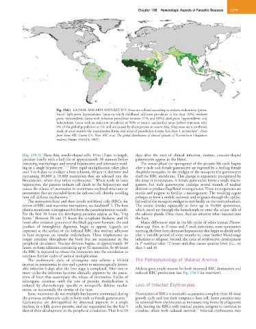
Fig. 158.1 GLOBAL MALARIA ENDEMICITY. Areas are colored according to malaria endemicity (preva-
lence): light green, hypoendemic (areas in which childhood infection prevalence is less than 10%); medium
green, mesoendemic (areas with infection prevalence between 11% and 50%); dark green, hyperendemic and
holoendemic (areas with an infection prevalence of 50% or more); unclassified areas (yellow) represent only
6% of the global population at risk and are caused by discrepancies in recent data. Gray areas are a combined
2
mask of areas outside the transmission limits and areas of population density less than 1 person/km . (Data
from Snow RW, Guerra CA, Noor AM, et al: The global distribution of clinical episodes of Plasmodium falciparum
malaria. Nature 434:214, 2005.)
(Fig. 158.2). These thin, needle-shaped cells, 10 to 12 µm in length, days after the start of clinical infection, mature, crescent-shaped
circulate briefly with a half-life of approximately 30 minutes before gametocytes appear in the blood.
traversing macrophages and several hepatocytes and ultimately resid- The sexual phase (or sporogony) of the parasite life cycle begins
ing in a single hepatocyte. 11,12 Here rapid multiplication takes place after a male and female gametocyte are ingested by a feeding female
over 5 to 8 days to produce a liver schizont, 80 µm in diameter and Anopheles mosquito. In the midgut of the mosquito the gametocytes
containing 30,000 ± 10,000 merozoites that are released into the shed the RBC membrane. This change is apparently precipitated by
13
bloodstream, where they infect erythrocytes. When ready to leave the drop in temperature. A female gametocyte forms a single macro-
hepatocytes, the parasite induces cell death in the hepatocytes and gamete, but male gametocytes undergo several rounds of nuclear
causes the release of merozoites in membrane-enclosed structures or division to produce flagellated microgametes. These microgametes are
merosomes that are extruded from the infected cell, thereby avoiding motile and migrate to fertilize a macrogamete. The resulting zygote
host cell defense mechanisms. 14 enlarges to form a mobile ookinete and migrates through the epithe-
The merozoites bind and then invade red blood cells (RBCs; for lial wall of the mosquito midgut to rest finally on the external surface.
15
review of RBC and merozoite interactions, see Satchwell ). The host The oocyst divides repeatedly to form up to 10,000 sporozoites,
plasma membrane is invaginated to form the parasitophorous vacuole. which travel up through the hemolymph to enter the acinar cells of
For the first 10 hours the developing parasites appear as fine “ring the salivary glands. Once there, they are infective when injected into
forms.” Between 10 and 15 hours the cytoplasm thickens, and 16 the host.
hours after invasion, granules of the black pigment hematin, the end Major differences exist in the life cycles of other human Plasmo-
product of hemoglobin digestion, begin to appear. Ligands are dium spp. First, in P. vivax and P. ovale infections, some sporozoites
expressed at the surface of the infected RBC that mediate adhesion entering the liver form dormant hypnozoites that begin to divide only
to host receptors on venular endothelium. These trophozoites no after a variable period of some months to cause further blood-stage
longer circulate throughout the body but are sequestered in the infections or relapses. Second, the cycle of erythrocytic development
peripheral circulation. Nuclear division begins, at approximately 30 in P. malariae takes 72 hours and thus causes quartan fever (i.e., on
hours, to form schizonts containing up to 32 merozoites. At 48 hours days 1 and 4).
the RBC is ruptured to release the merozoites into the circulation to
continue further cycles of asexual multiplication.
The erythrocytic cycle of schizogony may achieve a 10-fold The Pathophysiology of Malarial Anemia
increase in parasitemia in vivo and a patent or microscopically detect-
able infection 6 days after the liver stage is completed. After two or Malaria gives ample reasons for both increased RBC destruction and
more cycles the infection becomes clinically apparent by the parox- reduced RBC production (see Fig. 158.3 for overview).
ysms of fever that accompany the release of merozoites. Cycles of
schizogony continue until the rate of parasite multiplication is
reduced by chemotherapy, specific or nonspecific defense mecha- Loss of Infected Erythrocytes
nisms, or occasionally the demise of the host.
Some merozoites do not multiply but become committed during Destruction of RBCs is inevitable as parasites complete their 48-hour
16
the previous erythrocytic cycle to form male or female gametocytes. growth cycle and lyse their temporary host cell. Some parasites may
Gametocytes are distinguished by dispersed pigment in a single be removed from erythrocytes as immature ring forms by phagocytic
nucleus, in a fully grown parasite, and are sequestered for the first 5 cells, leaving the RBCs with residual parasite antigens to continue to
17
days of their development in the peripheral circulation. Thus 8 to 10 circulate, albeit with reduced survival. Infected erythrocytes may