
Page 2601 - Hematology_ Basic Principles and Practice ( PDFDrive )
P. 2601
2314 Part XIII Consultative Hematology
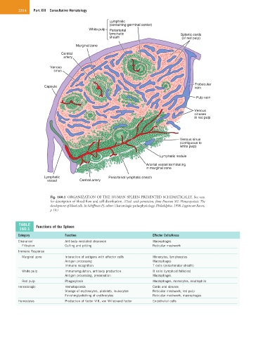
Lymphatic
(containing germinal center)
White pulp Periarterial
lymphatic Splenic cords
sheath (of red pulp)
Marginal zone
Central
artery
Venous
sinus
Trabecular
Capsule vein
Pulp vein
Venous
sinuses
in red pulp
Venous sinus
(contiguous to
white pulp)
Lymphatic nodule
Arterial vessel terminating
in marginal zone
Lymphatic Periarterial lymphatic sheath
vessel Central artery
Fig. 160.1 ORGANIZATION OF THE HUMAN SPLEEN PRESENTED SCHEMATICALLY. See text
for description of blood flow and cell distribution. (Used, with permission, from Emerson SG: Hematopoiesis: The
development of blood cells. In Schiffman FJ, editor: Hematologic pathophysiology, Philadelphia, 1998, Lippincott-Raven,
p 10.)
TABLE Functions of the Spleen
160.1
Category Function Effector Cells/Areas
Clearance/ Antibody-mediated clearance Macrophages
Filtration Culling and pitting Reticular meshwork
Immune Response
Marginal zone Interaction of antigens with effector cells Monocytes, lymphocytes
Antigen processing Macrophages
Immune recognition T cells (periarteriolar sheath)
White pulp Immunoregulation, antibody production B cells (lymphoid follicles)
Antigen processing, preservation Macrophages
Red pulp Phagocytosis Macrophages, monocytes, neutrophils
Hematologic Hematopoiesis Cords and sinuses
Storage of erythrocytes, platelets, leukocytes Reticular meshwork, red pulp
Finishing/polishing of erythrocytes Reticular meshwork, macrophages
Hemostasis Production of factor VIII, von Willebrand factor Endothelial cells