
Page 2602 - Hematology_ Basic Principles and Practice ( PDFDrive )
P. 2602
Chapter 160 The Spleen and Its Disorders 2315
Circumferential reticulum of
periarterial lymphatic sheath
Marginal zone Arterial vessel
Sinus
Sinus
Cord
Splenic cord Pulp vein
Trabecular
vein
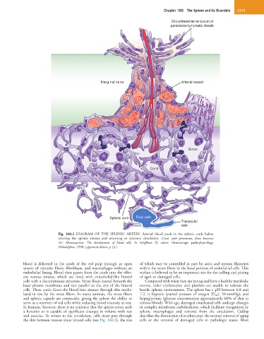
Fig. 160.2 DIAGRAM OF THE SPLENIC ARTERY. Arterial blood pools in the splenic cords before
entering the splenic sinuses and returning to systemic circulation. (Used, with permission, from Emerson
SG: Hematopoiesis: The development of blood cells. In Schiffman FJ, editor: Hematologic pathophysiology,
Philadelphia, 1998, Lippincott-Raven, p 12.)
blood is delivered to the cords of the red pulp through an open of which may be controlled in part by actin and myosin filaments
system of reticular fibers, fibroblasts, and macrophages without an within the stress fibers in the basal portion of endothelial cells. This
endothelial lining. Blood then passes from the cords into the effer- surface is believed to be an important site for the culling and pitting
ent venous sinuses, which are lined with endothelial-like littoral of aged or damaged cells.
cells with a discontinuous structure. Stress fibers extend beneath the Compared with when they are young and have a healthy metabolic
basal plasma membrane and run parallel to the axis of the littoral reserve, older erythrocytes and platelets are unable to tolerate the
cells. These cords direct the blood into sinuses through slits modu- hostile splenic environment. The spleen has a pH between 6.8 and
lated in size by the stress fibers. In many animals, the stress fibers 7.2, is hypoxic (partial pressure of oxygen [P O2]: 54 mmHg), and
and splenic capsule are contractile, giving the spleen the ability to hypoglycemic (glucose concentration approximately 60% of that in
serve as a reservoir of red cells while reducing blood viscosity at rest. venous blood). With age, damaged enucleated cells undergo changes
In humans, however, there is no evidence that the spleen serves such in complex membrane carbohydrates, which facilitate recognition by
a function or is capable of significant changes in volume with rest splenic macrophages and removal from the circulation. Culling
and exercise. To return to the circulation, cells must pass through describes the destruction of erythrocytes: the normal removal of aging
the slits between venous sinus littoral cells (see Fig. 160.3), the size cells or the removal of damaged cells in pathologic states. Most