
Page 281 - Hematology_ Basic Principles and Practice ( PDFDrive )
P. 281
232 Part III Immunologic Basis of Hematology
CTL CTL CTL
Perforin
granzyme
Perforin
Granzyme
Target cell Target cell
Target cell
Apoptosis
+
+
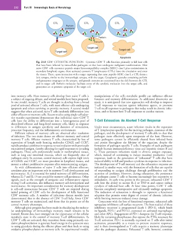
Fig. 21.8 CD8 CYTOLYTIC FUNCTION. Cytotoxic CD8 T cells function primarily to kill host cells
that have been infected by intracellular pathogens or that have undergone malignant transformation. After
+
naive CD8 cells encounter peptide–major histocompatibility complex (MHC) class I plus costimulation in
secondary lymphoid organs, these activated cytotoxic T lymphocytes (CTLs) leave the circulation and enter
the tissues. There, upon interaction with a target expressing that same peptide–MHC class I, a CTL forms a
lytic synapse, similar to the immunologic synapse, with the target. Cytoplasmic granules containing perforin
and granzymes congregate at the synapse, and granule contents are exocytosed into the cleft between the CTL
and its target cell. Perforin molecules facilitate entry of the cytolytic molecules into the target cells, and
granzymes act to promote apoptosis of the target cell.
into memory cells. How memory cells develop from naive T cells is manipulations of the cell’s metabolic profile can influence effector
a subject of ongoing debate, and several models have been proposed. function and memory differentiation. As additional discoveries are
In one model, memory T cells are thought to develop from a broad made, it is anticipated that new approaches will develop to improve
pool of activated effector T cells, with most effector cells undergoing T-cell responses to vaccines against infectious agents, to promote
apoptosis and others surviving to provide memory. A second model T-cell recall responses to pathogens that today result in chronic infec-
suggests that when activated, naive T cells randomly differentiate into tions, and to harness host T-cell responses to combat tumors.
either effectors or memory cells. Recent studies using single-cell adop-
+
tive transfer experiments demonstrate that individual naive CD8 T
cells have the ability to differentiate into a heterogeneous pool of T-Cell Exhaustion: An Aborted T-Cell Response
short-lived effector and long-lived memory cells, likely in response
to differences in antigen specificity and duration of stimulation, Under most circumstances, acute infection results in the expansion
precursor frequency, and the inflammatory environment. of T lymphocytes specific for the inciting pathogen, clearance of the
Different subsets of memory cells are observed after resolution pathogen, and the development of memory T cells able to clear that
of infection. The two main classes are effector memory and central pathogen more effectively upon reexposure of the host. However,
memory T cells. Effector memory T cells, characterized by loss of some pathogens cannot be efficiently cleared from infected hosts
expression of lymph node homing molecules CD62L and CCR7, and persist throughout the lifetime of the organism, despite the
rapidly produce cytokines in response to restimulation with previously formation of pathogen-specific T cells. Examples of such pathogens
encountered antigen, thereby allowing for rapid responses to invading include human immunodeficiency virus and hepatitis viruses B and
pathogens. These cells preferentially reside in nonlymphoid tissues, C. These persistent infections result in chronic antigen exposure,
such as lung and intestinal mucosa, which are frequently sites of which, instead of continuing to induce maximal productive T-cell
pathogen entry. In contrast, central memory cells express high levels responses, leads to the generation of “exhausted” T cells that have
of CD62L and CCR7, are more prevalent in lymphoid tissues, and reduced ability to kill and produce cytokines in response to infection.
mount a robust proliferative response after reencountering antigen. The development of T-cell memory and the exhaustion response are
As with differentiation of naive T cells into efficient effectors, initiated in similar ways, with the formation of cells that are capable
cytokines play an important role in memory T-cell development and of responding to antigen challenge through proliferation and the
maintenance. IL-2 is essential for initial memory cell differentiation, secretion of cytokines. However, during exhaustion, the persistence
whereas IL-7 and IL-15 are crucial for memory cell persistence. Other of pathogen causes T cells to become increasingly less responsive to
+
signals, such as the strength of antigenic and inflammatory signals stimulation. At early time points in this process, exhausted CD8 T
during T-cell activation, also influence memory cell development and cells lose the ability to secrete IL-2 or TNF-α and cannot induce
+
maintenance. An important consideration for memory development cytolysis of infected host cells. At later time points, CD8 T cells
+
is cell–cell interactions because CD4 T cells are required during become completely unresponsive and ultimately undergo apoptosis.
+
initial priming of CD8 cells for development of fully functional The induction of exhaustion is throught to represent a functional
+
CD8 memory cells. A number of infectious disease models have adaption that permits some degree of control of chronic infection
+
+
demonstrated that in the absence of CD4 T-cell help, fewer CD8 while limiting immune-induced tissue damage.
memory T cells are maintained, and those that do persist are of the Concurrent with the loss of functional responses, exhausted cells
central memory phenotype. upregulate inhibitory cell surface receptors. The best studied of these
Although great progress has been made in elucidating the molecu- inhibitory receptors is programmed death 1 (PD-1), which binds
lar underpinnings of immunologic memory, much remains to be its ligands, PD-L1 and PD-L2, expressed on activated macrophages
learned. Recent data have emerged on the importance of the cellular and other APCs. Engagement of PD-1 dampens the T-cell response,
metabolic state in the control of memory T-cell differentiation. As likely by recruiting phosphatases that oppose the PTKs necessary for
+
CD8 T cells are activated, they transition from using primarily oxi- T-cell activation. PD-1 is normally expressed on T cells after initial
dative phosphorylation to generate basal energy in the quiescent state activation, presumably as a means to prevent excessive responses,
to using glycolysis during the effector phase and then back to using and is then downregulated as T cells acquire a memory phenotype
oxidative phosphorylation as memory cells. In experimental models, after the pathogen clearance. Exhausted T cells, however, continue