
Page 283 - Hematology_ Basic Principles and Practice ( PDFDrive )
P. 283
234 Part III Immunologic Basis of Hematology
Cytokine
deprivation
CTLA4 CD28 TCR PD1 TGF-R
IL-2 Effector
cell
Inhibitory cytokines Phosphorylation-dependent
CD25 Inhibitory proximal signals Inhibitory
signals signals
IL-10
TGF-β DGKs
Shp-1
Effector Treg
cell
PLCγ1
DAG E3
PA PIP2
Ras
Apoptosis via IP3 Ub
cell-mediated contact PI3K
Fig. 21.9 T-REGULATORY CELL ACTIONS. T-regulatory cells (Tregs)
act to suppress other T cells through a multitude of mechanisms, including
the secretion of suppressor cytokines interleukin 10 (IL-10) and transforming Proteosomal
growth factor β (TGF-β), consumption of local concentrations of IL-2, and degradation
induction of cell cycle arrest or apoptosis.
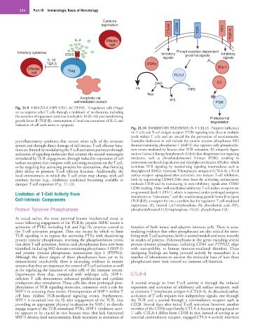
Fig. 21.10 INHIBITORY PATHWAYS IN T CELLS. Negative influences
on T cells and T-cell antigen receptor (TCR) signaling take place at multiple
levels within T cells and are crucial for the prevention of autoimmunity.
proinflammatory cytokines that recruit other cells of the immune Examples (indicated in red) include the protein tyrosine phosphatase SH2
system and through direct damage of self-tissues. T-cell effector func- domain-containing phosphatase-1 (SHP-1) that opposes early phosphoryla-
tions are limited by modulating the T-cell activation pathways through tion events mediated by kinases after TCR activation, E3 ubiquitin ligases
activation of signaling molecules that counter the second messengers such as Casitas b-lineage lymphoma-b (Cbl-b) that ubiquitinate key signaling
stimulated by TCR engagement, through inducible expression of cell mediators, such as phosphatidylinositol 3-kinase (PI3K), resulting in
surface receptors that compete with activating receptors on the T cell, proteosome-mediated degradation and diacylglycerol kinases (DGKs), which
or by targeting key activating proteins for destruction, thus limiting terminate TCR signaling by metabolizing signaling intermediates such as
their ability to promote T-cell effector function. Additionally, the diacylglycerol (DAG). Cytotoxic T-lymphocyte antigen-4 (CTLA-4), a T-cell
local environment in which the T cell exists may change, with cell surface receptor upregulated after activation, also induces T-cell inhibition,
extrinsic factors (e.g., inhibitory cytokines) becoming available to both by sequestering CD80/CD86 away from the activating costimulatory
dampen T-cell responses (Fig. 21.10). molecule CD28 and by transducing its own inhibitory signals after CD80/
CD86 binding. Other well-established inhibitory T-cell surface receptors are
Limitation of T-Cell Activity From programmed death 1 (PD-1), which is expressed under prolonged antigenic
stimulation or “exhaustion,” and the transforming growth factor β receptor
Cell-Intrinsic Components (TGF-β-R), a receptor for one a cytokine key for regulatory T cell–mediated
suppression. IP 3 , Inositol 1,4,5-trisphosphate; PA, phosphatidic acid; PIP 2 ,
Protein Tyrosine Phosphatases phosphatidylinositol-(4,5)-bisphosphate; PLCγ1, phospholipase Cγ1.
As noted earlier, the most proximal known biochemical event to
occur following engagement of the TCR by peptide–MHC results is
activation of PTKs, including Lck and Zap-70, enzymes central to function of both innate and adaptive immune cells. There is accu-
the T-cell activation program. Thus one means by which to limit mulating evidence that other phosphatases are also critical for inter-
TCR signaling is to oppose the activating PTKs with deactivating fering with T-cell activation, both in animal models and more recently
protein tyrosine phosphatases, reversing the phosphorylation events in studies of patients. Polymorphisms in the genes encoding several
that drive T-cell activation. Several such phosphatases have now been protein tyrosine phosphatases, including CD45 and PTPN22, align
identified, including SH2 domain-containing phosphatase 1 (SHP-1) with susceptibility to human immune-mediated disorders. These
and protein tyrosine phosphatase, nonreceptor type 1 (PTPN1). intriguing findings are being pursued actively by researchers in a
Although the direct targets of these phosphatases have yet to be number of laboratories to uncover the molecular basis of how these
demonstrated conclusively, there is increasing evidence in murine phosphatases exert their control on immune cell function.
systems that they are important for control of T-cell activation as well
as for regulating the function of other cells of the immune system.
Experiments show that, compared with wild-type cells, SHP-1- CTLA-4
deficient T cells demonstrate enhanced proliferation and cytokine
production after stimulation. These cells also show prolonged phos- A second strategy to limit T-cell activity is through the induced
phorylation of TCR signaling molecules, consistent with a role for expression and activation of inhibitory cell surface receptors, such
SHP-1 in reversing these events. Overexpression of SHP-1 within T as cytotoxic T lymphocyte antigen-4 (CTLA-4). As discussed earlier,
cell lines inhibits TCR-mediated signaling events. Furthermore, activation of T cells requires two independent signals, one through
SHP-1 is recruited into the IS after engagement of the TCR, thus the TCR and a second through a costimulatory receptor such as
providing an appropriate physical localization for SHP-1 to directly CD28. Several days after initial T-cell activation, however, another
engage targets of the TCR-stimulated PTKs. SHP-1 inhibitory activ- member of the CD28 superfamily, CTLA-4, becomes upregulated on
ity appears to be crucial in vivo because mice that lack functional T cells. CTLA-4 differs from CD28 in that, instead of serving as an
SHP-1 develop fatal autoimmunity, likely secondary to alterations of essential costimulatory receptor, engaged CTLA-4 actively interferes