
Page 278 - Hematology_ Basic Principles and Practice ( PDFDrive )
P. 278
Chapter 21 T-Cell Immunity 229
Consequences of
uncontrolled activity
Th1
IFN-γ Inflammation
T-bet
+IFN-γ,
IL-12 Th2
IL-4
+IL-4, TSLP, GATA3 IL-5 Allergy
IL-25, IL-33 IL-13
Naive +IL-6,
CD4 + IL-21, TGF-β Th17
IL-17a
RORγt Autoimmunity
+IL-21 IL-17f
Tfh
Bcl-6 IL-21 ?
+
+
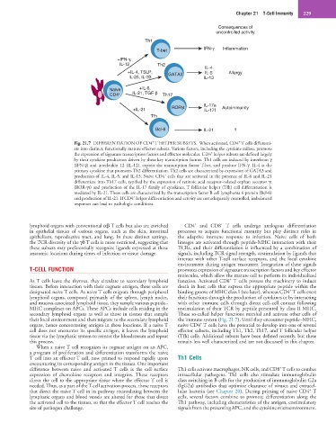
Fig. 21.7 DIFFERENTIATION OF CD4 T HELPER SUBSETS. When activated, CD4 T cells differenti-
ate into distinct, functionally mature effector subsets. Various factors, including the cytokine milieu, promote
+
the expression of signature transcription factors and effector molecules. CD4 helper subsets are defined largely
by their cytokine production driven by these key transcription factors. Th1 cells are induced by interferon γ
(IFN-γ) and interleukin 12 (IL-12), express the transcription factor T-bet, and produce IFN-γ. IL-4 is the
primary cytokine that promotes Th2 differentiation. Th2 cells are characterized by expression of GATA3 and
+
production of IL-4, IL-5, and IL-13. Naive CD4 cells that are activated in the presence of IL-6 and IL-21
differentiate into Th17 cells, typified by the expression of retinoic acid receptor–related orphan receptor γt
(ROR-γt) and production of the IL-17 family of cytokines. T follicular helper (Tfh) cell differentiation is
mediated by IL-21. These cells are characterized by the transcription factor B cell lymphoma 6 protein (Bcl-6)
+
and production of IL-21. If CD4 helper differentiation and activity are not adequately controlled, imbalanced
responses can lead to pathologic conditions.
+
+
lymphoid organs with conventional αβ T cells but also are enriched CD4 and CD8 T cells undergo analogous differentiation
in epithelial tissues of various organs, such as the skin, intestinal processes to acquire functional maturity but play distinct roles in
epithelium, reproductive tract, and lung. In these distinct settings, the adaptive immune response to infection. Naive cells of both
the TCR diversity of the γδ T cells is more restricted, suggesting that lineages are activated through peptide-MHC interaction with their
these subsets may preferentially recognize ligands expressed at these TCRs, and their differentiation is influenced by a combination of
anatomic locations during times of infection or tissue damage. signals, including TCR signal strength, costimulation by ligands that
interact with other T-cell surface receptors, and the local cytokine
environment during antigen encounter. Integration of these signals
T-CELL FUNCTION promotes expression of signature transcription factors and key effector
molecules, which allow the mature cell to perform its individualized
+
As T cells leave the thymus, they circulate to secondary lymphoid function. Activated CD8 T cells possess the machinery to induce
tissues. Before interaction with their cognate antigen, these cells are death in host cells that express the appropriate peptide within the
+
designated naive T cells. As naive T cells migrate through peripheral binding groove of MHC class I (see later), whereas CD4 T cells exert
lymphoid organs, composed primarily of the spleen, lymph nodes, their functions through the production of cytokines or by interacting
and mucosa-associated lymphoid tissue, they sample various peptide– with other immune cells through direct cell–cell contact following
MHC complexes on APCs. These APCs include cells residing in the restimulation of their TCR by peptide presented by class II MHC.
secondary lymphoid organs as well as those in tissues that sample These so-called helper functions marshal and activate other cells of
their local environment and then migrate to the secondary lymphoid the immune system (Fig. 21.7). Until they encounter peptide–MHC,
+
organs, hence concentrating antigen in these locations. If a naive T naive CD4 T cells have the potential to develop into one of several
cell does not encounter its specific antigen, it leaves the lymphoid effector subsets, including Th1, Th2, Th17, and T follicular helper
tissue via the lymphatic system to reenter the bloodstream and repeat (Tfh) cells. Additional subsets have been defined recently, but these
this process. remain less well characterized and are not discussed in this chapter.
When a naive T cell recognizes its cognate antigen on an APC,
a program of proliferation and differentiation transforms the naive
T cell into an effector T cell, now primed to respond rapidly upon Th1 Cells
encountering its corresponding antigen in the tissues. One important
+
difference between naive and activated T cells is the cell surface Th1 cells activate macrophages, NK cells, and CD8 T cells to combat
expression of chemokine receptors and integrins. These receptors intracellular pathogens. Th1 cells also stimulate immunoglobulin
direct the cell to the appropriate tissue where the effector T cell is class switching in B cells for the production of immunoglobulin G2a
needed. Thus, as a part of the T-cell activation process, those receptors (IgG2a) antibodies that optimize clearance of viruses and extracel-
+
that direct the naive T cell in its pathway recirculating between the lular bacteria (see Chapter 20). During priming of naive CD4 T
lymphatic organs and blood vessels are altered for those that direct cells, several factors combine to promote differentiation along the
the activated cell to the tissues, so that the effector T cell reaches the Th1 pathway, including characteristics of the antigen, costimulatory
site of pathogen challenge. signals from the presenting APC, and the cytokine microenvironment.