
Page 311 - Hematology_ Basic Principles and Practice ( PDFDrive )
P. 311
Chapter 23 Dendritic Cell Biology 259
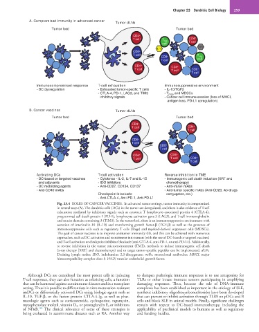
A. Compromised immunity in advanced cancer Tumor dLNs
Tumor bed Tumor bed
CD4 +
T cell T reg CD4 +
cell T cell
TGFβ
IL-10
CD8 +
T cell + T reg
CD8
T cell cell
CD4 + CD4 +
T cell T cell CD8 +
T cell
Immunocompromised response T-cell exhaustion Immunosuppressive environment
- DC dysregulation - Exhausted tumor-specific T cells - IL-10/TGFβ
- CTLA-4, PD-1, LAG3, and TIM3 - T regs and MDSCs
inhibitory signals - Cancer cell immune evasion (loss of MHCI,
antigen loss, PD-L1 upregulation)
B. Cancer vaccines Tumor dLNs
Tumor bed Tumor bed
CD4 +
T cell CD4 +
T cell
CD8 +
T cell
CD8 +
T cell
CD4 + CD4 +
T cell T cell CD8 +
T cell
Activating DCs T-cell activation Reverse inhibition in TME
- DC-based or targeted vaccines - Cytokines - IL-2, IL-7 and IL-15 - Immunogenic cell death induction (XRT and
and adjuvants - IDO inhibitors chemotherapy)
- DC mobilizing agents - Anti-CD27, CD134, CD137 - Anti-VEGF mAbs
- Anti-CD40 mAbs - Anti-tumor specific mAbs (Anti-CD20, Ab-drugs
Checkpoint blockade congugates, etc.)
- Anti-CTLA-4, Anti-PD-1, Anti-PD-L1
Fig. 23.4 ROLES OF CANCER VACCINES. In advanced tumor settings, tumor immunity is compromised
in several ways (A). The dendritic cells (DCs) in the tumor are deregulated, and there is also evidence of T-cell
exhaustion mediated by inhibitory signals such as cytotoxic T lymphocyte–associated protein 4 (CTLA-4),
programmed cell death protein 1 (PD-1), lymphocyte activation gene 3 (LAG3), and T-cell immunoglobulin
and mucin domain containing-3 (TIM3). In the tumor bed, there is an immunosuppressive environment with
secretion of interleukin-10 (IL-10) and transforming growth factor-β (TGF-β) as well as the presence of
immunosuppressive cells such as regulatory T cells (Tregs) and myeloid-derived suppressor cells (MDSCs).
The goal of cancer vaccines is to improve antitumor immunity (B), and this can be achieved with numerous
approaches, such as DC activation and recruitment into tumors (with the use of DC-based or targeted vaccines)
and T-cell activation or checkpoint inhibitor blockade (anti-CTLA-4, anti-PD-1, or anti-PD-L1). Additionally,
to reverse inhibition in the tumor microenvironment (TME), methods to induce immunogenic cell death
(x-ray therapy [XRT] and chemotherapy) and to target tumor-specific peptides can be implemented. dLNs,
Draining lymph nodes; IDO, indoleamine 2,3-dioxygenase; mAbs, monoclonal antibodies; MHCI, major
histocompatibility complex class I; VEGF, vascular endothelial growth factor.
Although DCs are considered the most potent cells in inducing to dampen pathologic immune responses is to use antagonists for
T-cell responses, they can also function as tolerizing cells, a function TLRs or other innate immune sensors participating in amplifying
that can be harnessed against autoimmune diseases and in a transplant damaging responses. Thus, because the role of DNA–immune
setting. Thus it is possible to differentiate in vitro maturation-resistant complexes has been established as important in the etiology of SLE,
imDCs or differentially activated DCs using biologic agents such as synthetic inhibitory oligodeoxyribonucleotides have been developed
IL-10, TGF-β, or the fusion protein CTLA-4-Ig, as well as phar- that can prevent or inhibit activation through TLR9 on pDCs and B
macologic agents such as corticosteroids, cyclosporine, rapamycin, cells and block SLE in animal models. Finally, significant challenges
mycophenolate mofetil, vitamin D 3, or prostaglandin E 2 or inhibitors remain with respect to DC-based immunotherapy, including the
184
of NFκB. The clinical relevance of some of these strategies is applicability of preclinical models to humans as well as regulatory
being evaluated in autoimmune diseases such as RA. Another way and funding hurdles.