
Page 307 - Hematology_ Basic Principles and Practice ( PDFDrive )
P. 307
Chapter 23 Dendritic Cell Biology 255
Inhibition of viral replication
Death receptor NK cell activation
ligands Cross-priming
Enhancement of primary antibody response
Membrane Isotope switching
PRR
IFN-α Naive CD8
Cytoplasmic PRR
Cytotoxicity
TNF-α
pDC Antigen Naive CD4
Intemalization
Bacteria, viruses, Infection Maturation presentation Treg
“danger signals”
activation
TNF-α Mature DC TGF-β
Th17
Immature DC
IL-6 Th1
IL-12 IFN-γ
cytotoxicity
NK cell
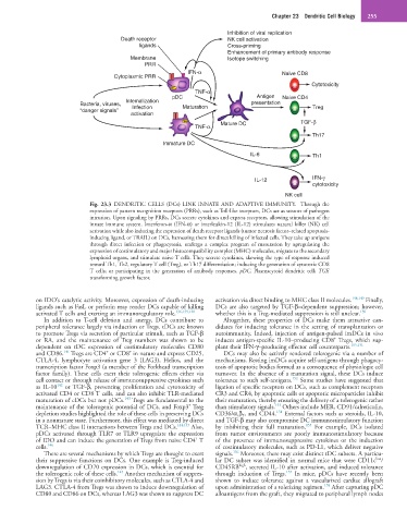
Fig. 23.3 DENDRITIC CELLS (DCs) LINK INNATE AND ADAPTIVE IMMUNITY. Through the
expression of pattern recognition receptors (PRRs), such as Toll-like receptors, DCs act as sensors of pathogen
intrusion. Upon signaling by PRRs, DCs secrete cytokines and express receptors, allowing stimulation of the
innate immune system. Interferon-α (IFN-α) or interleukin-12 (IL-12) stimulates natural killer (NK) cell
activation while also inducing the expression of death receptor ligands (tumor necrosis factor–related apoptosis-
inducing ligand, or TRAIL) on DCs, harnessing them for direct killing of infected cells. They take up antigens
through direct infection or phagocytosis, undergo a complex program of maturation by upregulating the
expression of costimulatory and major histocompatibility complex (MHC) molecules, migrate to the secondary
lymphoid organs, and stimulate naive T cells. They secrete cytokines, skewing the type of response induced
toward Th1, Th2, regulatory T cell (Treg), or Th17 differentiation; inducing the generation of cytotoxic CD8
T cells; or participating in the generation of antibody responses. pDC, Plasmacytoid dendritic cell; TGF,
transforming growth factor.
on IDO’s catalytic activity. Moreover, expression of death-inducing activation via direct binding to MHC class II molecules. 148,149 Finally,
ligands such as FasL or perforin may render DCs capable of killing DCs are also targeted by TGF-β–dependent suppression; however,
activated T cells and exerting an immunoregulatory role. 136,139,140 whether this is a Treg-mediated suppression is still unclear. 150
In addition to T-cell deletion and anergy, DCs contribute to Altogether, these properties of DCs make them attractive can-
peripheral tolerance largely via induction or Tregs. tDCs are known didates for inducing tolerance in the setting of transplantation or
to promote Tregs via secretion of particular stimuli, such as TGF-β autoimmunity. Indeed, injection of antigen-pulsed imDCs in vivo
+
or RA, and the maintenance of Treg numbers was shown to be induces antigen-specific IL-10–producing CD8 Tregs, which sup-
dependent on tDC expression of costimulatory molecules CD80 plant their IFN-γ–producing effector cell counterparts. 105,151
+
+
141
and CD86. Tregs are CD4 or CD8 in nature and express CD25, DCs may also be actively rendered tolerogenic via a number of
CTLA-4, lymphocyte activation gene 3 (LAG3), Helios, and the mechanisms. Resting imDCs acquire self-antigens through phagocy-
transcription factor Foxp3 (a member of the Forkhead transcription tosis of apoptotic bodies formed as a consequence of physiologic cell
factor family). These cells exert their tolerogenic effects either via turnover. In the absence of a maturation signal, these DCs induce
152
cell contact or through release of immunosuppressive cytokines such tolerance to such self-antigens. Some studies have suggested that
142
as IL-10 or TGF-β, preventing proliferation and cytotoxicity of ligation of specific receptors on DCs, such as complement receptors
activated CD4 or CD8 T cells, and can also inhibit TLR-mediated CR3 and CR4, by apoptotic cells or apoptotic microparticles inhibit
143
maturation of cDCs but not pDCs. Tregs are fundamental to the their maturation, thereby ensuring the delivery of a tolerogenic rather
+
153
maintenance of the tolerogenic potential of DCs, and Foxp3 Treg than stimulatory signals. Others include MER, CD91/calreticulin,
154
depletion studies highlighted the role of these cells in preserving DCs CD36/α v β 5 , and CD44. External factors such as steroids, IL-10,
in a nonmature state. Furthermore, this effect was mediated by direct and TGF-β may also compromise DC immunostimulatory function
155
TCR–MHC class II interactions between Tregs and DCs. 144,145 Also, by inhibiting their full maturation. For example, DCs isolated
pDCs activated through TLR7 or TLR9 upregulate the expression from tumor environments are poorly immunostimulatory because
+
of IDO and can induce the generation of Tregs from naive CD4 T of the presence of immunosuppressive cytokines or the induction
cells. 146 of costimulatory molecules, such as PD-L1, which deliver negative
156
There are several mechanisms by which Tregs are thought to exert signals. Moreover, there may exist distinct tDC subsets. A particu-
low
their suppressive functions on DCs. One example is Treg-induced lar DC subset was identified in normal mice that were CD11c /
downregulation of CD70 expression in DCs, which is essential for CD45RB high , secreted IL-10 after activation, and induced tolerance
147
157
the tolerogenic role of these cells. Another mechanism of suppres- through induction of Tregs. In mice, pDCs have recently been
sion by Tregs is via their coinhibitory molecules, such as CTLA-4 and shown to induce tolerance against a vascularized cardiac allograft
158
LAG3. CTLA-4 from Tregs was shown to induce downregulation of upon administration of a tolerizing regimen. After capturing pDC
CD80 and CD86 on DCs, whereas LAG3 was shown to suppress DC alloantigens from the graft, they migrated to peripheral lymph nodes