
Page 449 - Hematology_ Basic Principles and Practice ( PDFDrive )
P. 449
370 Part IV Disorders of Hematopoietic Cell Development
Passive transplacental passage of IgG antiplatelet antibodies into
fetal circulation can cause rapid destruction of fetal platelets. This
occurs in two circumstances: a (1) maternal autoimmune disease
such as idiopathic thrombocytopenic purpura or systemic lupus
erythematosus and (2) in neonatal alloimmune thrombocytopenia
by alloimmunization of the pregnant mother to fetal antigens inher-
ited from father but absent in the mother. In the former situation,
the mother has thrombocytopenia or a history of such; in the latter
situation, the mother has a normal platelet count and serum antibod-
ies to human platelet alloantigens.
Thrombocytopenia with absent radii syndrome is distinguished
from CAMT because in TAR, the radii are absent. Peripheral blood
chromosomes analysis is not associated with increased breakage with
DEB or MMC clastogenic stress testing, which allows CAMT to be
distinguished from FA. Increased platelet destruction also occurs in
newborns with giant benign hemangiomas of skin, liver, or spleen,
the so-called Kasabach-Merritt syndrome.
In an infant or young child with a CAMT clinical diagnosis but
without mutant MPL, other inherited forms of thrombocytopenia
should be addressed (see Table 29.5). These can generally be classified
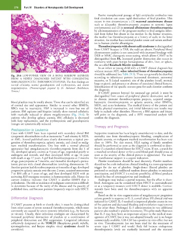
Fig. 29.6 LOW-POWER VIEW OF A BONE MARROW ASPIRATE according to inheritance pattern (autosomal dominant, autosomal
FROM A NEWLY DIAGNOSED PATIENT WITH CONGENITAL recessive, or X-linked recessive), size of the platelets (small, normal,
AMEGAKARYOCYTIC THROMBOCYTOPENIA. The three findings are large or giant), and presence or absence of associated clinical features.
normal cellularity, normal granulopoiesis and erythropoiesis, and absent Identification of the specific mutant gene for each disorder confirms
megakaryocytes. (Photomicrograph prepared by Dr. Mohamed Abdelhaleem, the diagnosis.
Toronto.)
If CAMT presents beyond the neonatal age period, it must be
distinguished from causes of peripheral platelet destruction such as
in chronic immune thrombocytopenia purpura, acquired amega-
blood platelets may be totally absent. Those that can be identified are karyocytic thrombocytopenia or aplastic anemia, other IBMFSs,
of normal size and appearance. Similar to several other IBMFSs, MDS, and acute leukemias. The medical history of the patient and
RBCs may be macrocytic. HbF is increased in most but not all family, physical examination, and initial laboratory test results may
patients. BM aspirates and biopsies initially show normal cellularity help to exclude other disorders. However, a BM aspirate and biopsy
with markedly reduced or absent megakaryocytes (Fig. 29.6). In will point to the diagnosis, and a MPL mutational analysis will
patients who develop aplastic anemia, BM cellularity is decreased confirm the diagnosis.
with fatty replacement, and the erythropoietic and granulopoietic
lineages are symmetrically reduced.
Therapy and Prognosis
Predisposition to Leukemia
Cases with CAMT have been reported with secondary clonal BM Supportive treatment has been largely unsatisfactory to date, and the
cytogenetic abnormalities such as monosomy 7 and trisomy 8, MDS, mortality rate from thrombocytopenic bleeding, complications of
or AML. Several published cases clearly demonstrate a typical pro- aplastic anemia, or malignant myeloid transformation has been very
gression of thrombocytopenia, aplastic anemia, and clonal or malig- close to 100%. For that reason, HLA typing of family members
nant myeloid transformation. One boy with a normal physical should be performed as soon as the diagnosis is confirmed to deter-
appearance had amegakaryocytic thrombocytopenia from day 1 of mine if a matched related donor for HSCT exists. If not, a search for
life, developed aplastic anemia at 5 years of age, responded poorly to a matched unrelated donor or for a cord blood graft should ensue as
androgens and steroids, and then developed AML at age 16 years soon as the severity of the clinical picture is appreciated. The need
with death at age 17 years. A girl had thrombocytopenia at 2 months for transfusional support is a cogent indication.
of age, pancytopenia at 5 months, and thereafter developed a preleu- Platelet transfusions should be used discretely. Platelet numbers
kemic picture with clonal abnormalities involving chromosome 19. should not be a sole indication; clinical bleeding is a more appropriate
Another patient had thrombocytopenia at 6 months of age, developed trigger for the use of platelets. Single-donor filtered platelets are
progressive aplastic anemia over the next 2 years, acquired monosomy preferred to multiple unfiltered random donor platelets to minimize
7 in BM cells at 5 years of age, and then developed MDS with an sensitization, and if HSCT is a realistic possibility, all blood products
activating RAS oncogene mutation in hematopoietic cells. Hence the should be free of cytomegalovirus and irradiated.
current evidence indicates that CAMT is another IBMFS that is Androgens may induce a partial response, but the effect is short-
preleukemic. The risk or incidence of malignant conversion is difficult lived. Androgens can be considered when HSCT is contraindicated
to determine because of the rarity of the disease and the paucity of or as a temporary measure until HSCT donor is available. Cortico-
published data, and because patients frequently require early HSCT. steroids have been used for thrombocytopenia with no apparent
efficacy.
Based on the in vitro augmentation of megakaryocyte progenitor
Differential Diagnosis colony growth in response to IL-3, a small phase I/II clinical trial was
initiated for CAMT. IL-3 resulted in improved platelet counts in two
If CAMT presents at birth or shortly after, it must be distinguished of five patients and decreased bleeding and transfusion requirements
from other causes of severe neonatal thrombocytopenia, which most in the other three. Prolonged IL-3 administration in two additional
commonly are caused by severe systemic infections (e.g., by bacteria patients also resulted in platelet increments. This pilot study illustrates
or viruses). Usually, these infectious etiologies are characterized by that IL-3 may have been an important adjunct to the medical man-
increased peripheral destruction of platelets or a combination of agement of CAMT, but it was not adopted broadly and is no longer
peripheral destruction and BM suppression. Congenital infections commercially available. GM-CSF has a positive in vitro effect but not
collectively designated as the TORCH (Toxoplasma gondii, rubella, in vivo. Thrombopoietin has not been tried for the treatment of
cytomegalovirus, and herpes simplex virus) syndrome should be severe type I CAMT and would likely fail because endogenous
considered. thrombopoietin levels are markedly increased and the mutated