
Page 453 - Hematology_ Basic Principles and Practice ( PDFDrive )
P. 453
374 Part IV Disorders of Hematopoietic Cell Development
previously suspected and that was only unmasked by testing in long- Clinical Features
term cultures. This observation, however, is in keeping with the
clinical observation that in addition to anemia, patients may have DBA registries with longitudinal data and a summary of published
neutropenia and thrombocytopenia. These findings were extended cases provide comprehensive information about clinical aspects of the
with evidence in long-term culture initiating assays for a trilineage disorder. Aside from findings associated with anemia, about half of
defect in DBA refractory to treatment. The data broaden the defini- infants at presentation look healthy and are normal physically. Unless
tion of DBA and explain generalized BM dysfunction and hypoplasia the baby develops cardiac failure as a result of anemia, hepatospleno-
in some cases of DBA that have puzzled investigators for years. megaly and edema are absent.
The molecular mechanism that links ribosome protein haploinsuf- Pregnancy, birth history, or both are often abnormal. In a survey
ficiency to the erythroid defect is unclear. One hypothesis is that it is from the French and German DBA registries of 64 pregnancies in 26
related to translation insufficiency. It is well known that during early women with DBA, complications were seen in 42 pregnancies (66%)
stages of erythropoiesis, translation is increased. It is possible that the and included abortion, preeclampsia, in utero fetal death, in utero
need for protein synthesis is not met during this critical developmental growth retardation, retroplacental hematoma, and preterm delivery.
stage. A second hypothesis is that ribosomal protein gene mutations Thirteen of 34 children born alive had DBA. Fetal DBA with hydrops
lead to accumulation of abnormal rRNA precursors as well as dysregu- fetalis has been reported. In current reports, more than 90% of cases
lation of multiple ribosomal protein genes and protein expression as present in the first 12 months of life; however, because of the avail-
shown with RPS19. This leads to defective ribosome biogenesis, ability of genetic testing, patients with mild to moderate phenotype
unassembled ribosome proteins, and cellular stress. Indeed, loss of are diagnosed later on in life. After diagnosis, family screening may
ribosome proteins have been shown to increase the levels of S6 kinase identify the parents or older siblings as affected.
phosphorylation via increased ROS, which in turn results in stimulat- About 30% to 47% of patients present with one or more con-
ing erythroid cell autophagy. A third hypothesis and the one consid- genital anomalies. Most of these phenotypic abnormalities belong to
ered most plausible is that defective ribosome biogenesis leads to the following categories: (1) craniofacial dysmorphism, including
activation of p53, thereby causing apoptosis and cell cycle arrest. A hypertelorism, microcephaly, microphthalmos, congenital cataract or
role of p53 is supported by a recent mouse model with mutations in glaucoma, strabismus, microretrognathism, and a high-arched palate
RPS19 that is characterized by RBC underproduction and small or cleft palate; (2) prenatal or postnatal growth failure independent
mouse size and by zebrafish models of RPS19 inhibition that manifest of steroid therapy; (3) neck anomalies, which may consist of a pte-
impaired erythropoiesis and malformations. Activation of p53 may rygium coli or the fusion of cervical vertebrae with flaring of the
involve the interactions of MDM2 with specific ribosomal proteins trapezius muscle (Klippel-Feil syndrome), giving a Turner syndrome
such as RPL5, RPL11, and RPL23. These interactions may lead to appearance or there may also be the Sprengel deformity (congenital
dissociation of p53 from MDM2, impairment of p53 targeting to the elevation of the scapula) as an isolated anomaly or a combination of
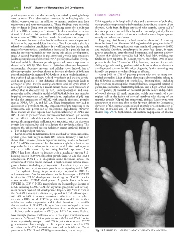
proteosome, and prevention of proteosome degradation. However, the two anomalies; and (4) thumb malformations, such as bifid
these models do not explain how haploinsufficiency of RPL5 and thumb (Fig. 29.7), duplication, subluxation, hypoplasia, or absence
RPL11 leads to p53 activation. Further, coinhibition of Tp53 activity
in five different zebrafish models of ribosome protein knockdown
rescued the morphologic malformations associated with the ribosome
protein knockdown, but did not alleviate the erythroid aplasia. This
suggests that ribosomal protein deficiency causes erythroid failure in
a Tp53-independent manner.
Extraribosomal functions have been ascribed to various ribosomal
protein genes that might mediate BM failure. Recently it has been
shown that ribosome protein haploinsufficiency results in decreased
GATA1 mRNA translation. This observation might be at least in part
responsible for the erythropoietic defect as the defective erythropoiesis
can be partially rescued by increasing GATA1 expression. Also,
RPS19 has been shown to interact with a nucleolar protein S19-
binding protein (S19BP), fibroblast growth factor 2, and the PIM-1
oncoprotein. PIM-1 is a ubiquitous serine-threonine kinase, the
expression of which can be induced in erythropoietic cells by several
growth factors, including erythropoietin. Thus there may be a pos-
sible link between erythropoietic growth factor signaling and RPS19.
The erythroid lineage is predominantly impaired in DBA for
unknown reasons. Studies have shown that the heme exporter FLVCR1
is critical for CFU-E development. Knocking out FLVCR1 in mice
causes impaired CFU-E development. A partial block in human
+
FLVCR1 in CD34 HSCs recapitulates the hematologic features of
+
+
DBA, including CD36 /CD135a erythroid progenitor cell develop-
ment but not myeloid cell development. Importantly, 55% to 95% of
the FLVCR1 transcript is alternatively spliced in DBA cells compared
with 4% to 24% in normal immature erythroid cells. The spliced
variants in DBA encode FLVCR1 proteins that are defective in their
cellular and surface expression and in their function. It is possible
that expression of FLVCR1 splicing variants leads to impaired export
of intracellular iron and apoptosis because of accumulation of iron.
Patients with mutations in RPL5 and RPL11 are more likely to
have multiple physical malformations. For example, thumb anomalies
are seen in 56% and 39% of patients with RPL5 and RPL11 muta-
tions, respectively, compared with 7% in patients who have RPS19
gene mutations. Interestingly, cleft lip or palate was reported in 42%
of patients with RPL5 mutations compared with 6% and 0% of
patients with RPL11 and RPS19 gene mutations, respectively. Fig. 29.7 BIFID THUMB IN DIAMOND-BLACKFAN ANEMIA.