
Page 454 - Hematology_ Basic Principles and Practice ( PDFDrive )
P. 454
Chapter 29 Inherited Bone Marrow Failure Syndromes 375
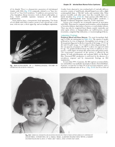
of the thumb. There is a characteristic association of triphalangeal Another facies observed in two unrelated girls of markedly different
thumbs with DBA (Fig. 29.8) commonly referred to as “Aase syn- ancestries consists of small heads, almond-shaped eyes with a slight
drome II” or “Aase-Smith syndrome.” In addition, some patients have antimongoloid slant, a “fish-like” smile, and a pointed chin. These
a flat, hypoplastic thenar eminence, weak or absent radial pulses, or patients resemble each other more than they resemble their own
both, which probably represent variations of the thumb family members (Fig. 29.9A–B). Some patients with DBA have a
malformations. phenotype indistinguishable from Treacher-Collins syndrome, a
Some patients have a characteristic facial appearance. The facies disorder of ribosome biogenesis caused by TCOF1 mutations.
of individuals with DBA is said to consist of tow-colored hair, a snub Various other anomalies are occasionally reported in association
nose, wide-set eyes, a thick upper lip, and an intelligent expression. with DBA. There may be urogenital malformations, such as dysplastic
or horseshoe kidneys, duplication of ureters, or renal tubular acidosis.
There may also be congenital heart disease, mainly ventricular and
atrial septal defects, or hypogonadism, ear malformations, mental
retardation, congenital hip dislocation, or tracheoesophageal fistula.
Laboratory Findings
Peripheral Blood and Bone Marrow. The main hematologic find-
ings in DBA are summarized in Table 29.6. The anemia is usually
profound at the time of diagnosis. Hemoglobin levels average 6.5 g/
dL in patients diagnosed in the first 2 months of life (range, 1.7–9.1 g/
dL) and 4.0 g/dL (range, 1.8–7.4 g/dL) in those diagnosed later. In
the vast majority of patients, the MCV is above the expected values
for age. The peripheral blood smear may show, in addition to mac-
rocytes, a mild degree of nonspecific anisocytosis and poikilocytosis.
The aregenerative component of the anemia is reflected by the
absence of both polychromasia and nucleated RBCs on the blood
film. Decreased RBC production is confirmed by the absence of a
reticulocyte response and by characteristic findings on BM
examination.
In more than 90% of patients, the BM aspirate is normocellular,
but erythroblasts are markedly decreased or absent. Proerythroblasts,
Fig. 29.8 RADIOGRAPH OF A TRIPHALANGEAL THUMB IN if present, account for less than 3% of all nucleated elements, with a
DIAMOND-BLACKFAN ANEMIA. myeloid-to-erythroid ratio of 10 to 1 (Fig. 29.10). In 5% to 10% of
A B
Fig. 29.9 SIMILAR DIAMOND-BLACKFAN FACIES IN TWO UNRELATED GIRLS OF DIFFERENT
ANCESTRIES, CONSISTING OF A SMALL HEAD, ALMOND-SHAPED EYES WITH A SLIGHT
ANTIMONGOLOID SLANT, A “FISH-LIKE” SMILE, AND A POINTED CHIN.