
Page 496 - Hematology_ Basic Principles and Practice ( PDFDrive )
P. 496
416 Part IV Disorders of Hematopoietic Cell Development
Acetylcholinesterase from erythrocytes and alkaline phosphatase these cell surface GPI-anchored proteins are manifold; they can serve
from leukocytes were the first GPI-anchored proteins shown to be as complement regulatory proteins, enzymes, blood group antigens,
missing in PNH. Since then, more than a dozen GPI-anchored receptors, and adhesion molecules. Membrane inhibitor of reactive
proteins with heterogeneous expression on hematopoietic cells have lysis (CD59) and decay accelerating factor (CD55)—both comple-
been found to be missing in PNH (Table 31.1). The functions of ment regulatory proteins—are the most widely expressed GPI-
anchored proteins and can be found on all hematopoietic lineages
−
+
including CD34 CD38 stem/progenitor cells. Certain proteins,
Monoclonal antibody CD58 (LFA3) and CD16 (FcγRIII), may exist in both GPI-linked
and transmembrane forms.
Protein
Phosphatidylinositol-Glycan Complementation
Proaerolysin Class A Gene
FLAER
Ethanolamine
p Investigators in Osaka, Japan, first identified the gene that was defec-
Glycan Glucosamine tive in PNH. The gene was isolated by expression cloning and named
p PIGA. PIGA was then cloned into an expression vector and trans-
Ethanolamine fected into GPI-deficient cell lines derived from PNH patients; cell
Inositol surface expression of all the missing GPI-anchored proteins was
restored, confirming that PIGA mutations are responsible for causing
P PNH. Since this seminal discovery, somatic mutations of the PIGA
PIPLC gene have been found in virtually all PNH patients to date. Little to
no GPI anchor is made when the PIGA gene is mutated. Conse-
quently, the translated protein (e.g., CD59, CD55, etc.) residing in
Plasma the cisterna of the endoplasmic reticulum cannot be attached to the
membrane GPI anchor and is degraded in situ.
The human PIGA gene contains six exons, five introns, and
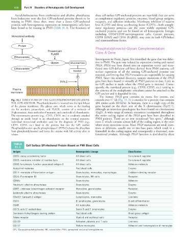
Fig. 31.1 STRUCTURE OF THE GLYCOSYLPHOSPHATIDYLINOSI- extends over 17 kb (Fig. 31.2); it encodes for a protein that contains
TOL (GPI) ANCHOR. Phosphatidylinositol is inserted into the lipid bilayer 484 amino acids (60 kDa). In humans, there is a single copy of the
of the plasma membrane. The glycan core, which serves as the binding gene located on the short arm of the X chromosome (Xp22.1),
site for aerolysin, proaerolysin, and FLAER, consists of a molecule of although an intronless pseudogene has been found on chromosome
N-glucosamine, three molecules of mannose, and a molecule of ethanolamine. 12q21. A wide range of somatic mutations interspersed throughout
The representative protein (e.g., CD55, CD59, etc.) is covalently attached the entire coding region of the PIGA gene have been described in
through an amide bond to an ethanolamine on the terminal mannose. PNH patients. There are no true mutational “hot spots,” although
Individual monoclonal antibodies used for the diagnosis of PNH (e.g., exon 2, which contains almost half of the coding region, is the exon
CD55, CD59, etc.) bind to the protein, but not the GPI anchor. where most mutations occur. Most PIGA mutations are small inser-
Phosphatidylinositol-specific phospholipase C (PIPLC) cleaves the phosphate tions or deletions, usually one or two base pairs, which result in a
from phosphatidylinositol and leaves the enzyme with full activity after its frameshift in the coding region and consequently a shortened, non-
release. functional product. Although PIGA function is abolished by these
TABLE Cell Surface GPI-Anchored Protein Absent on PNH Blood Cells
31.1
Antigen Hematopoietic Lineage Classification
CD55: decay accelerating factor All blood cells Complement regulator
CD59: membrane inhibitor of reactive lysis All blood cells Complement regulator
CD58: lymphocyte function associated antigen-3 All blood cells Adhesion molecule
Acetylcholinesterase Red blood cells Enzyme
CD14: monocyte differentiation antigen Granulocytes, monocytes, macrophages Endotoxin-binding receptor
CD16: Fcγ receptor III Granulocytes, NK cells Receptor
CD66b Granulocytes Adhesion
Neutrophil alkaline phosphatase Granulocytes Enzyme
CD87: urokinase (plasminogen activator) receptor Monocytes, granulocytes Receptor
Leukocyte alkaline phosphatase Granulocytes Enzyme
CDw52: Campath-1 antigen Lymphocytes, monocytes Unknown
CD24 B lymphocytes, granulocytes B-cell differentiation
CD48 All leukocytes Adhesion molecule
CD73: ecto-5′-nucleotidase Some B and T lymphocytes Enzyme
Dombrock-Holley/Gregory-bearing protein Red blood cells Blood group antigen
Folate receptor Myeloid and erythroid cells Receptor
CD109 Activated platelets and T cells Unknown
CD157 Mature monocytes Adhesion and transmigration of monocytes
GPI, Glycosylphosphatidylinositol; NK, natural killer; PNH, paroxysmal nocturnal hemoglobinuria.