
Page 574 - Hematology_ Basic Principles and Practice ( PDFDrive )
P. 574
Chapter 36 Disorders of Iron Homeostasis 489
phenotypical and genotypical screening should lead to a definitive Timing of Chelation Therapy
diagnosis in most patients. Aceruloplasminemia is a rare disorder, but
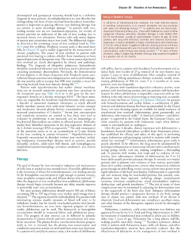
distinguishing this form of iron overload from hereditary hemochro- In all forms of transfusional iron overload, the most effective means
matosis is important in guiding effective iron-chelating therapy that of avoiding complications is to prevent excessive iron accumulation
24
can prevent or arrest neurologic damage. In patients with iron- with early iron-chelating therapy. 16,26 In patients who are transfusion-
loading anemia who are not transfusion-dependent, the severity of dependent from early infancy (i.e., those with thalassemia major or other
anemia provides no indication of the risk of iron loading due to congenital refractory anemias), chelation therapy is best started after
increased dietary iron absorption. Patients with only minor degrees 10–20 transfusions, usually at approximately 3 years of age. In older
of anemia may accumulate major iron loads. The differential diagno- patients with acquired refractory anemias who become transfusion-
sis directed at the remaining causes of iron overload listed in Table dependent, it seems advisable to begin chelation early after transfusion
36.3 poses few problems. Porphyria cutanea tarda is discussed more of 10–20 units of blood. In patients with iron-loading anemia and those
with sickle cell disease who are chronically transfused for prevention of
fully in Chapter 38 and is readily diagnosed by the measurement of complications, early therapy also seems prudent. 16,26 In each of these
urinary porphyrins. The source of iron overload in patients with disorders, delay in beginning chelation therapy only exposes the patient
parenteral iron loading is evident, whether from transfusion or from to a greater risk of iron toxicity.
repeated injections of therapeutic iron. The various causes of perinatal
iron overload are clearly distinguished by clinical and pathologic
findings. The diagnosis of idiopathic pulmonary hemosiderosis
should be considered whenever iron-deficiency anemia develops with will suffice, but in a patient with hereditary hemochromatosis and an
coexisting pulmonary abnormalities. Previously, the demonstration initial body iron burden of 25 g, removal of the iron burden may
of iron deposits in the brain of patients with Friedreich ataxia, pan- require 2 years or more of phlebotomy. After complete removal of
tothenate kinase-associated neurodegeneration, and neuroferritinopa- the iron load, lifelong maintenance therapy is needed, usually neces-
thy was possible only at autopsy, but MRI now provides a means for sitating phlebotomy of 500 mL every 3 to 4 months or, in some
detecting localized brain iron deposits during life. 20,24 patients, even less frequently.
Patients with hyperferritinemia but neither clinical manifesta- For patients with transfusion-dependent refractory anemia, most
tions nor an elevated transferrin saturation may have mutations in patients with iron-loading anemia, and rare patients with hemochro-
the ferroportin gene (see Table 36.4) or in the gene for the iron- matosis for whom phlebotomy is impossible, treatment with an iron
30
responsive element in L-ferritin messenger RNA. The latter muta- chelator is the only means of preventing or removing toxic accumula-
tions are responsible for hereditary hyperferritinemia with cataract, tions of iron (see box on Timing of Chelation Therapy). In patients
a disorder of autosomal dominant inheritance in which affected with hemochromatosis and cardiac failure, a combination of phle-
family members present with early-onset bilateral nuclear cataracts botomy and chelation therapy has been recommended. In the United
and moderately elevated plasma ferritin concentrations caused by States, two iron-chelating agents are available for initial treatment of
30
increased concentrations of L-ferritin. Serum iron concentration transfusional iron overload: deferoxamine, given parenterally, and
16
and transferrin saturation are normal or low, body iron level as deferasirox, administered orally. A third iron chelator—oral deferi-
evaluated by phlebotomy is not increased, and no hematologic or prone—is approved in the United States, the European Union, and
biochemical abnormalities are evident in affected persons. Molecular other countries for patients with thalassemia major when deferox-
studies have identified mutations in the iron-responsive element of amine is contraindicated or inadequate.
30
the L-ferritin messenger RNA as responsible. The only consequence Over the past 4 decades, clinical experience with deferoxamine, a
of the mutation seems to be an accumulation of L-type ferritin hexadentate bacterial siderophore purified from Streptomyces pilosus,
30
in the lens, resulting in cataract formation. Hyperferritinemia is has established the efficacy and safety of this agent in preventing
frequently encountered in disorders without iron overload, includ- organ dysfunction and prolonging survival in patients with transfu-
ing malignancy, rheumatologic diseases (such as systemic juvenile sional iron overload. 16,26 Unfortunately, deferoxamine given orally is
idiopathic arthritis, adult-onset Still disease, and hemophagocytic poorly absorbed. To be effective, the drug must be administered by
lymphohistiocytosis/macrophage activation syndrome), and chronic prolonged subcutaneous or intravenous infusion with a small portable
infection. syringe pump, ideally each day, making compliance a demanding
task. In patients with modest iron loads and no evidence of iron
toxicity, slow subcutaneous infusion of deferoxamine for 9 to 12
Therapy hours daily usually provides adequate therapy. In severely iron-loaded
patients and in patients with evidence of iron toxicity, particularly
The goal of therapy for iron overload is reduction and maintenance those with cardiac complications, chronic slow intravenous infusions
of body iron at normal or near-normal levels. If possible, phlebotomy given through an indwelling central venous catheter may permit more
is the treatment of choice for hemochromatosis, iron-loading anemia rapid reduction of the body iron burden. Deferoxamine is a generally
(if the hemoglobin concentration is high enough to permit venesec- safe and nontoxic drug for iron-loaded patients, but systemic com-
tion), porphyria cutanea tarda, and African dietary iron overload. 14,15 plications have been reported, including allergic anaphylactoid
After the diagnosis of iron overload has been established, phlebotomy reactions, infectious complications, visual abnormalities, auditory
therapy should begin promptly because any delay extends exposure dysfunction, and growth retardation. 16,26 The risk of many of these
to potentially toxic iron accumulations. complications may be minimized by adjusting the deferoxamine dose
For most patients, phlebotomy should remove 500 mL of blood, to the magnitude of the body iron load. Adequate deferoxamine
containing 200 to 250 mg of iron, once weekly, until storage iron is therapy should produce a progressive decrease in the body storage
depleted. 14,15 The regimen should be individualized. For patients with iron of almost any patient with iron overload. If no decline is
iron-loading anemia, smaller amounts of blood will need to be observed, blood and deferoxamine use, compliance, ascorbate status,
withdrawn weekly, but for heavily iron-loaded patients with heredi- and other features of the therapeutic regimen should be thoroughly
tary hemochromatosis, an even more vigorous program of twice- reassessed.
weekly phlebotomy can be used. The hematocrit or hemoglobin Deferasirox, a synthetic, orally active tridentate iron chelator, was
concentration should be measured before each phlebotomy proce- approved for use by the U.S. Food and Drug Administration in 2005
dure. The progress of iron removal can be followed by periodic for treatment of transfusional iron overload in adults and in children
measurements of plasma ferritin and iron concentrations and trans- older than 2 years of age. Deferasirox has a long plasma half-life,
ferrin saturation. The plasma ferritin concentration declines progres- making possible once-daily dosing. 16,26 Extensive systematic clinical
sively as iron is removed, but the plasma iron concentration and trials in patients with thalassemia major, sickle cell disease, and other
transferrin saturation remain elevated until iron stores near depletion. transfusion-dependent anemias have provided evidence that the
In a patient with porphyria cutanea tarda, a few weeks of phlebotomy effectiveness of deferasirox in the management of iron overload is