
Page 572 - Hematology_ Basic Principles and Practice ( PDFDrive )
P. 572
Chapter 36 Disorders of Iron Homeostasis 487
5th decade of life with a triad of diabetes mellitus, progressive neu-
rologic disease (dementia, dysarthria, and dystonia), and retinal
degeneration.
Patients with autosomal dominant hemochromatosis resulting
from mutations in the ferroportin gene that compromise iron export,
such as those resulting in ferroportins that are unable to reach the
cell surface to interact with hepcidin, have iron deposition predomi-
nantly in macrophages, are almost devoid of clinical manifestations,
18
and apparently do not require treatment. Patients with mutations
that result in ferroportins that reach the cell surface but do not
respond to hepcidin develop a parenchymal pattern of iron overload
that resembles that found in patients with the autosomal recessive
forms of hemochromatosis. 18
Acquired Iron Overload
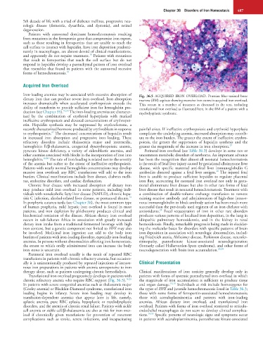
Iron-loading anemias may be associated with excessive absorption of Fig. 36.5 ACQUIRED IRON OVERLOAD. Prussian blue–stained bone
dietary iron that can produce severe iron overload. Iron absorption marrow (BM) aspirate showing excessive iron stores in acquired iron overload.
increases dramatically when accelerated erythropoiesis exceeds the This occurs in a number of instances as discussed in the text, including
ability of transferrin to provide sufficient iron for hemoglobin pro- transfusional iron overload as illustrated here, in the BM of a patient with a
duction (see Chapter 35). 16,26 The iron-loading anemias are character- myelodysplastic syndrome.
ized by the combination of erythroid hyperplasia with marked
ineffective erythropoiesis and elevated concentrations of erythropoi-
etin. Hepcidin synthesis may be suppressed by erythroferrone, a
recently characterized hormone produced by erythroblasts in response painful crises. If ineffective erythropoiesis and erythroid hyperplasia
27
to erythropoietin. The decreased concentrations of hepcidin result complicate the underlying anemia, increased absorption may contrib-
in increased iron absorption and progressive iron loading. These ute to the iron burden. The greater the extent of ineffective erythro-
refractory disorders include thalassemia major and intermedia, poiesis, the greater the suppression of hepcidin synthesis and the
hemoglobin E/β-thalassemia, congenital dyserythropoietic anemia, greater the magnitude of the increase in iron absorption. 27
pyruvate kinase deficiency, a variety of sideroblastic anemias, and Perinatal iron overload (see Table 36.3) develops in some rare or
other anemias associated with blocks in the incorporation of iron into uncommon metabolic disorders of newborns. An important advance
hemoglobin. 16,26 The rate of iron loading is related not to the severity has been the recognition that almost all neonatal hemochromatosis
of the anemia but rather to the extent of ineffective erythropoiesis. is the result of fetal liver injury caused by gestational alloimmune liver
Patients with nearly normal hemoglobin concentrations may develop disease, with specific maternal anti-fetal liver immunoglobulin G
28
massive iron overload; any RBC transfusions will add to the iron antibodies directed against a fetal liver antigen. The injured fetal
burden. Clinical manifestations include liver disease, diabetes melli- liver is unable to produce sufficient hepcidin to regulate placental
tus, endocrine disorders, and cardiac dysfunction. iron flux, accounting for neonatal iron overload not only in gesta-
Chronic liver disease with increased absorption of dietary iron tional alloimmune liver disease but also in other rare forms of fetal
may produce mild iron overload in some patients, including indi- liver disease that result in neonatal hemochromatosis. Treatment with
viduals with nonalcoholic fatty liver disease (NAFLD), chronic hepa- a combination of double-volume exchange transfusion to remove
14
titis C infection, alcohol-related liver disease, or portacaval shunts. existing reactive antibody and administration of high-dose intrave-
In porphyria cutanea tarda (see Chapter 38), the most common type nous immunoglobulin to block antibody action has been much more
of human porphyria, mild hepatic iron overload is found in most successful than the previously used regimen of an iron chelator with
28
patients, and iron depletion by phlebotomy produces clinical and antioxidants. Focal sequestration of iron in other rare disorders
biochemical remission of the disease. African dietary iron overload produces various patterns of localized iron deposition, in the lung in
occurs in sub-Saharan Africa in association with greatly increased idiopathic pulmonary hemosiderosis, and in the kidney in renal
dietary iron intake from a traditional fermented beverage with high hemosiderosis. Finally, remarkable progress is being made in elucidat-
iron content, but a genetic component not linked to HFE may also ing the molecular bases for disorders with specific patterns of brain
be involved. Medicinal iron ingestion can add to the body iron iron deposition in association with neurologic abnormalities, includ-
burden of patients with iron-loading disorders, especially iron-loading ing Friedreich ataxia, Alzheimer disease, Parkinson disease, neurofer-
anemias. In persons without abnormalities affecting iron homeostasis, ritinopathy, pantothenate kinase-associated neurodegeneration
the extent to which orally administered iron can increase the body (formerly called Hallervorden-Spatz syndrome), and other forms of
iron stores is uncertain. neurodegeneration with brain iron accumulation. 20,24
Parenteral iron overload usually is the result of repeated RBC
transfusions in patients with chronic refractory anemia, but occasion-
ally it is unintentionally produced by repeated injections of intrave- Clinical Presentation
nous iron preparations in patients with anemia unresponsive to iron
therapy alone, such as patients undergoing chronic hemodialysis. Clinical manifestations of iron toxicity generally develop only in
Transfusional iron overload progressively develops in patients with patients with forms of systemic parenchymal iron overload in which
chronic refractory anemia who require RBC support (Fig. 36.5). 16,26 the magnitude of iron accumulation is sufficient to produce tissue
In patients with severe congenital anemias such as thalassemia major and organ damage. 14,18 Individuals at risk include homozygotes for
(Cooley anemia) or Blackfan-Diamond syndrome, transfusional iron the types of HFE and juvenile hemochromatosis listed in Table 36.3;
loading begins in infancy. Severe iron loading may develop in those with some forms of ferroportin-associated hemochromatosis;
transfusion-dependent anemias that appear later in life, namely, those with aceruloplasminemia; and patients with iron-loading
aplastic anemia, pure RBC aplasia, hypoplastic or myelodysplastic anemias, African dietary iron overload, and transfusional iron
disorders, and the anemia of chronic renal failure. Patients with sickle overload. Patients with forms of iron overload restricted to reticulo-
cell anemia or sickle cell/β-thalassemia are also at risk for iron over- endothelial macrophages do not seem to develop clinical complica-
load if chronically given transfusions for prevention of recurrent tions. 14,18 Specific patterns of neurologic signs and symptoms occur
complications such as stroke, severe infections, and incapacitating in patients with aceruloplasminemia, pantothenate kinase-associated