
Page 570 - Hematology_ Basic Principles and Practice ( PDFDrive )
P. 570
Chapter 36 Disorders of Iron Homeostasis 485
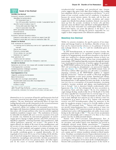
TABLE Causes of Iron Overload reticuloendothelial macrophage and parenchymal sites. Genetic
36.3 studies suggest that genes other than those leading to iron loading
23
Hereditary Iron Overload have substantial effects on iron accumulation and toxicity. In other
forms of iron overload, another level of complexity is introduced
Autosomal recessive hemochromatosis because the central nervous system, the testes, and the fetus are
Hereditary hemochromatosis functionally separate from the systemic circulation and cannot
HFE-associated (type 1) acquire iron directly from plasma transferrin. Instead, iron must be
Non-HFE-associated: transferrin receptor 2-associated (type 3) taken up from the systemic circulation by barrier cells and then
Juvenile hemochromatosis (type 2) exported across the blood–brain and blood–cerebrospinal fluid bar-
Hemojuvelin-associated (type 2A) riers into the brain interstitial and cerebrospinal fluids, across the
Hepcidin-associated (type 2B) blood–testis barrier, and across the placenta to the fetus. As a
24
DMT1-associated hemochromatosis consequence, disorders affecting the proteins responsible for iron
Atransferrinemia supply to these compartments have distinctive manifestations.
Aceruloplasminemia
Autosomal dominant hemochromatosis
Ferroportin-associated with impaired iron export (type 4A) Hereditary Iron Overload
Ferroportin-associated with hepcidin resistance (type 4B)
Acquired Iron Overload Within the systemic circulation, the specific patterns of iron depo-
From increased iron absorption sition and damage found in the hereditary disorders of iron over-
Iron-loading anemia (refractory anemia with hypercellular erythroid load can be characterized by reference to the pathways of internal
marrow) iron exchange shown in Fig. 35.1 and the classification given in
Chronic liver disease Table 36.4.
Porphyria cutanea tarda In HFE hemochromatosis, an autosomal recessive disorder, the
African dietary iron overload* underlying genetic defect in the regulation of hepcidin production
Medicinal iron ingestion* results in an inappropriately elevated iron absorption at any level of
From parenteral iron body iron, resulting in a chronic progressive increase in body iron
Transfusional iron overload stores along with enhanced release of iron from reticuloendothelial
Inadvertent iron overload from therapeutic injections macrophages. HFE regulates hepcidin expression through the hepatic
Perinatal Iron Overload bone morphogenetic protein/sons of mothers against decapentaple-
Gestational alloimmune liver disease with neonatal hemochromatosis gic (BMP/SMAD) pathway by binding to the BMP type I receptor
Trichohepatoenteric syndrome Alk3 and preventing its ubiquitination and proteasomal degrada-
25
Cerebrohepatorenal (Zellweger) syndrome tion (see Chapter 35). The C282Y and H63D mutant forms of
GRACILE (Fellman) syndrome HFE fail to stabilize Alk3 expression and cell surface accumulation,
Focal Sequestration of Iron impairing activation of the BMP/SMAD pathway to suppress
25
Idiopathic pulmonary hemosiderosis hepcidin production. Patients are unable to effectively upregulate
Renal hemosiderosis hepcidin expression as iron stores increase. Intestinal iron absorp-
Associated with neurologic abnormalities tion, although inappropriately high in hereditary HFE-associated
Pantothenate kinase-associated neurodegeneration hemochromatosis, is still regulated by body iron levels. As the body
Neuroferritinopathy iron level rises as a consequence of increased absorption, circulating
Friedreich ataxia transferrin becomes saturated and plasma non-transferrin-bound
21
iron is formed. Iron is deposited initially predominantly within
*May have a genetic component. hepatocytes (Fig. 36.4), but subsequently the iron accumulates in
DMT1, Divalent metal transporter 1; GRACILE, growth retardation, 21
aminoaciduria, cholestasis, iron overload, lactic acidosis, and early death. the pancreas, heart, and other organs. By the time symptoms of
organ damage develop, usually in the 4th or 5th decade of life, body
iron stores typically have increased from the normal range of 1 g or
less to 15 to 20 g or more. Further increments in body iron stores
abnormalities in the interaction of hepcidin and ferroportin that lead may be fatal, although some patients are able to tolerate a total iron
to excessive intestinal iron absorption, resulting in body iron accu- accumulation of as much as 40 to 50 g. Patients with autosomal
mulation. The rate, distribution, and harmful effects of tissue iron recessive non-HFE hemochromatosis caused by mutations in the
loading depend on the specific abnormality in the interaction between gene for transferrin receptor 2 seem to be clinically similar to those
hepcidin and ferroportin produced by each mutation. with the HFE-associated form. 14,18 Patients with autosomal reces-
In general, cellular iron loading in the autosomal recessive disor- sive juvenile hemochromatosis have a similar pattern of tissue iron
ders begins with formation of plasma nontransferrin-bound iron that deposition found in HFE hemochromatosis but develop severe iron
then enters cells through pathways other than the carefully regulated overload much earlier, with hypogonadism and cardiac disease mani-
21
transferrin–transferrin receptor route. The mechanisms of toxicity festing in the 2nd decade of life. 14,18 The rate of iron accumulation
in vulnerable iron-loaded cells seem to involve expansion of the pool is increased substantially and is estimated to be three to four times
of cytosolic iron followed by iron-induced generation of reactive greater than that in HFE-associated disease.
oxygen species; damage to lipids, proteins, and DNA; and injury to Patients with DMT1-associated hemochromatosis have in
subcellular organelles, including lysosomes and mitochondria, with common a severe microcytic anemia with low hepcidin, high transfer-
22
cellular dysfunction, apoptosis, and necrosis. The pattern of the rin saturation, and marked hepatic iron deposition but normal to
organs affected, the timing of the onset of toxic manifestations, and moderately elevated serum ferritin concentration. 14,18 Congenital
the severity of tissue damage are known to be influenced by a variety atransferrinemia (hypotransferrinemia) is a rare disorder of autosomal
of factors in both hereditary and acquired varieties of systemic iron recessive inheritance in which plasma transferrin is nearly absent and
overload. Within the systemic circulation, these factors include (1) hepcidin is decreased. 14,18 Patients have a severe hypochromic micro-
the specific underlying genetic or acquired abnormality; (2) the cytic anemia and die without transferrin infusion or blood transfu-
magnitude of iron excess; (3) the rate of iron loading; (4) the distribu- sions. Hereditary aceruloplasminemia (hypoceruloplasminemia) is a
tion of iron load among more innocuous storage deposits in reticu- rare disorder of iron homeostasis inherited as an autosomal recessive
loendothelial macrophages and potentially injurious accumulations trait, resulting from absence or severe deficiency of ceruloplasmin
24
in parenchymal cells of the liver, pancreas, heart, and other organs; occurring as a consequence of mutations in the ceruloplasmin gene.
and (5) the extent of internal redistribution of iron between Patients with aceruloplasminemia typically present in the fourth or