
Page 645 - Hematology_ Basic Principles and Practice ( PDFDrive )
P. 645
Chapter 40 Thalassemia Syndromes 547
either highly insoluble or form homotetramers (Hb H and Hb Bart) β-thalassemia alleles, one on each copy of chromosome 11. As a
that are incapable of releasing oxygen normally, and because they are consequence of diminished Hb A synthesis, the circulating RBCs are
relatively unstable, will precipitate as the cell ages. For poorly under- very hypochromic, abnormal in shape, and they contain markedly
stood reasons, no compensatory regulatory mechanism exists whereby reduced amounts of Hb. Accumulation of free α-globin chains leads
impaired synthesis of one globin subunit leads to a compensatory to the deposition of precipitated aggregates of these chains to the
downward adjustment in the production of the other (partner) globin detriment of the erythrocyte and its precursor cells in the bone
chain of the Hb tetramer. Thus, whereas useless excess α-globin marrow (BM). The anemia of thalassemia major is so severe that
chains continue to accumulate and precipitate in β-thalassemia, long-term blood transfusions are usually required for survival.
excess β-globin chains form Hb H in α-thalassemia. The abnormal The term β-thalassemia intermedia is applied to a less severe clini-
solubility or oxygen-carrying properties of these chains lead to a cal phenotype in which significant anemia occurs but chronic transfu-
variety of physiologic derangements. Indeed, in the severe forms of sion therapy is not absolutely required. It usually results from the
thalassemia, it is the behavior of the unpaired globin chains accumu- inheritance of two β-thalassemia mutations, one mild and one severe;
lating in relative excess that dominates the pathophysiology of the the inheritance of two mild mutations; or, occasionally, the inheri-
syndrome rather than the mere underproduction of functioning Hb tance of complex combinations, such as a single β-thalassemia defect
tetramers. The precise complications of this pathophysiologic phe- and an excess of normal α-globin genes, or two β-thalassemia muta-
nomenon are diverse and depend on the amount and the identity of tions coinherited with heterozygous α-thalassemia (in this last form,
the globin chain accumulating in excess. The fundamental principle known as αβ-thalassemia, the α-thalassemia allele reduces the burden
that must be appreciated is that thalassemias cause symptoms by of unpaired α-chains). 12–14 Simple heterozygosity for certain forms of
underproduction of Hb and by accumulation of unpaired globin β-thalassemic hemoglobinopathies can also be associated with a
subunits. The unpaired subunits are usually the major sources of thalassemia intermedia phenotype, sometimes called dominant
morbidity and mortality. β-thalassemia. 15,16
The predominant circulating Hb at the moment of birth is fetal Thalassemia minor, also known as β-thalassemia trait or hetero-
hemoglobin (Hb F α2γ2 ) (see Chapter 33). Although the switch from zygous β-thalassemia, is caused by the presence of a single
γ- to β-globin biosynthesis begins before birth, the composition of β-thalassemia mutation and a normal β-globin gene on the other
Hb in the peripheral blood changes much later because of the long- chromosome. It is characterized by profound microcytosis with
life span of normal circulating red blood cells (RBCs) (approximately hypochromia but mild or minimal anemia. In general, thalassemia
120 days). Hb F is thus slowly replaced by Hb A so that infants do minor has no associated symptoms, although cholelithiasis has been
not depend heavily on normal amounts and function of Hb A until reported from the accumulation of pigmented gallstones as a result
they are between 4 and 6 months old. The pathophysiologic conse- of hemolysis in this population. 17
quences of these considerations are that whereas α-chain hemoglo-
binopathies tend to be symptomatic in utero and at birth, individuals
with β-chain abnormalities are asymptomatic until 4 to 6 months of Molecular Pathology
age. These differences in the onset of phenotypic expression arise
because α-chains are needed to form Hb F and Hb A, but β-chains Forms of β-thalassemia arise from mutations that affect every step in
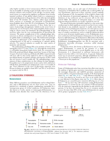
are required only for Hb A. the pathway of globin gene expression: transcription, processing of
the messenger ribonucleic acid (mRNA) precursor, translation of
mature mRNA, and posttranslational integrity of the β-polypeptide
β-THALASSEMIA SYNDROMES chain (Fig. 40.1 and Table 40.1). 18–20 Large deletions removing two
or more non–α-genes are found in rare cases, as are smaller partial
Nomenclature or total deletions of the β-gene alone (see Fig. 40.1). Most types of
β-thalassemia are caused by point mutations affecting one or a few
Many different mutations cause β-thalassemia and its related disor- bases. 18–25 Of the more than 200 mutations causing β-thalassemia,
ders, such as δβ-thalassemia and the silent carrier state. They are approximately 15 account for the vast majority of affected patients,
inherited in a multitude of genetic combinations responsible for a with the remainder responsible for the disorder in only relatively few
heterogeneous group of clinical syndromes. β-Thalassemia major, patients. It has been determined that five or six mutations usually
also known as Cooley anemia or homozygous β-thalassemia, is a account for more than 90% of the cases of β-thalassemia in a given
clinically severe disorder that results from the inheritance of two ethnic group or geographic area (see Table 40.1). 22
5 ′ 1 2 3 3 ′
β-Globin gene 100 bp
Transcription Frameshift RNA cleavage
RNA splicing Nonsense codon Initiator codon
Cap site Unstable globin Small deletion
Fig. 40.1 MODEL OF THE HUMAN β-GLOBIN GENE SHOWING SITES AND TYPES OF VARIOUS
MUTATIONS CAUSING β-THALASSEMIA. (Adapted from Kazazian HH Jr: The thalassemia syndromes:
molecular basis and prenatal diagnosis in 1990. Semin Hematol 27:209, 1990.)