
Page 646 - Hematology_ Basic Principles and Practice ( PDFDrive )
P. 646
548 Part V Red Blood Cells
TABLE Common β-Thalassemia Mutations in Different Racial boundaries for normal splicing and thereby completely block produc-
40.1 Groups tion of mature functional messenger RNA. Thus, no β-globin can be
synthesized (β°-thalassemia). Other mutations alter the consensus
Racial Group Description sequences that surround the GT- and AG-invariant dinucleotides and
Mediterranean IVS-1, position 110 (G → A) decrease the efficiency of normal splicing signals by 70% to 95%,
+
Codon 39, nonsense (CAG → TAG) resulting in β -thalassemia; some consensus mutations even abolish
IVS-1, position 1 (G → A) splicing completely, causing β°-thalassemia. A third type of splicing
IVS-2, position 745 (C → G) aberration results from mutations that are not in the immediate
IVS-1, position 6 (T → C) vicinity of a normal splice site. These alter regions within the gene,
IVS-2, position 1 (G → A) called cryptic splice sites, which resemble consensus splicing sites but
do not normally sustain splicing (see Fig. 40.2). The mutations
African –34 (A → G) activate the site by supplying a critical GT or AG nucleotide or by
–88 (C → T) creating a sufficiently strong consensus signal to stimulate splicing at
Poly(A), (AATAAA → AACAAA)
that site 60% to 100% of the time. The activated cryptic sites gener-
Southeast Asian Codons 41/42, frameshift (-CTTT) ate an abnormally spliced, untranslatable mRNA species. Only 10%
IVS-2, position 654 (C → T) to 40% of the mRNA precursors are thus spliced at the normal sites,
+
–28 (A → T) which causes β -thalassemia of variable severity. The mutation
Asian Indian IVS-1, position 5 (G → C) responsible for the most common form of β-thalassemia among
619-bp deletion Greeks and Cypriots (Fig. 40.3) activates a cryptic splice site near the
26,27
Codons 8/9, frameshift (++G) 3′ end of the first intron (position 110). The determinants that
Codons 41/42, frameshift (–CTTT) dictate the degree to which each mutation alters splice site use remain
IVS-1, position 1 (G → T) largely unknown.
Data from Kazazian HH Jr, Boehm CD: Molecular basis and prenatal diagnosis
of beta-thalassemia. Blood 72(4):1107, 1988; and Kazazian HH Jr, Boehm CD:
personal communication, 1993. Translation
Mutations that abolish translation occur at several locations along the
mature mRNA and are very common causes of β-thalassemia (see
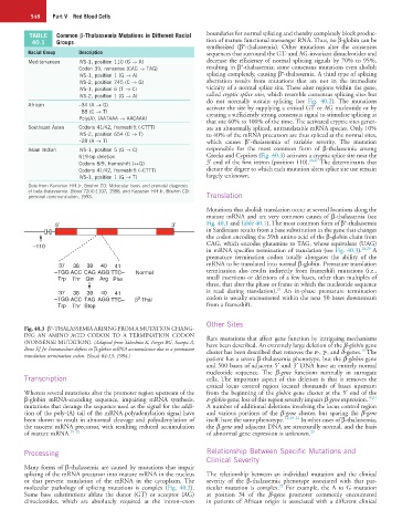
5 ′ 3 ′ Fig. 40.1 and Table 40.1). The most common form of β°-thalassemia
in Sardinians results from a base substitution in the gene that changes
the codon encoding the 39th amino acid of the β-globin chain from
–110 CAG, which encodes glutamine to TAG, whose equivalent (UAG)
28,29
in mRNA specifies termination of translation (see Fig. 40.3). A
premature termination codon totally abrogates the ability of the
37 38 39 40 41 mRNA to be translated into normal β-globin. Premature translation
–TGG ACC CAG AGG TTC– Normal termination also results indirectly from frameshift mutations (i.e.,
Trp Thr Gin Arg Phe small insertions or deletions of a few bases, other than multiples of
three, that alter the phase or frame in which the nucleotide sequence
29
37 38 39 40 41 is read during translation). An in-phase premature termination
0
–TGG ACC TAG AGG TTC– β Thal codon is usually encountered within the next 50 bases downstream
Trp Thr Stop from a frameshift.
Other Sites
Fig. 40.3 β°-THALASSEMIA ARISING FROM A MUTATION CHANG-
ING AN AMINO ACID CODON TO A TERMINATION CODON Rare mutations that affect gene function by intriguing mechanisms
(NONSENSE MUTATION). (Adapted from Takeshita K, Forget BG, Scarpa A, have been described. An extremely large deletion of the β-globin gene
Benz EJ Jr: Intranuclear defects in β-globin mRNA accumulation due to a premature cluster has been described that removes the ε-, γ-, and δ-genes. The
30
translation termination codon. Blood 64:13, 1984.)
patient has a severe β-thalassemia phenotype, but the β-globin gene
and 500 bases of adjacent 5′ and 3′ DNA have an entirely normal
nucleotide sequence. The β-gene functions normally in surrogate
Transcription cells. The important aspect of this deletion is that it removes the
critical locus control region located thousands of bases upstream
Whereas several mutations alter the promoter region upstream of the from the beginning of the globin gene cluster at the 5′ end of the
β-globin mRNA-encoding sequence, impairing mRNA synthesis, ε-globin gene; loss of this region severely impairs β-gene expression. 18,31
mutations that derange the sequence used as the signal for the addi- A number of additional deletions involving the locus control region
tion of the poly-(A) tail of the mRNA polyadenylation signal have and various portions of the β-gene cluster, but sparing the β-gene
been shown to result in abnormal cleavage and polyadenylation of itself, have the same phenotype. 1,2,19–21 In other cases of β-thalassemia,
the nascent mRNA precursor, with resulting reduced accumulation the β-gene and adjacent DNA are structurally normal, and the basis
of mature mRNA. 19–21 of abnormal gene expression is unknown. 22
Processing Relationship Between Specific Mutations and
Clinical Severity
Many forms of β-thalassemia are caused by mutations that impair
splicing of the mRNA precursor into mature mRNA in the nucleus The relationship between an individual mutation and the clinical
or that prevent translation of the mRNA in the cytoplasm. The severity of the β-thalassemia phenotype associated with that par-
22
molecular pathology of splicing mutations is complex (Fig. 40.2). ticular mutation is complex. For example, the A to G mutation
Some base substitutions ablate the donor (GT) or acceptor (AG) at position 34 of the β-gene promoter commonly encountered
dinucleotides, which are absolutely required at the intron–exon in patients of African origin is associated with a different clinical