
Page 647 - Hematology_ Basic Principles and Practice ( PDFDrive )
P. 647
Chapter 40 Thalassemia Syndromes 549
A mutation is inherited. The differences in physiologically impor-
Normal splicing tant functions among haplotypes that modulate severity remain
unknown, but a possible explanation lies in the variable abilities of
Splice Entire IVS-1 removed Splice the γ-globin genes on different chromosomes to respond to severe
erythroid stress by increased expression during postnatal life. The
GGC AG g t .. .. . . . .. . .c ctat t g g tc tattt tc cc a cc ct t ag G CTG β-globin genes carried on some haplotypes differ in the degree to
29 30 31 33
which they can respond in this manner. Because Hb F synthesis
1
...
ATG GTG CTG GGC AGG CTG CTG....... CAC TAA reduces the severity of β-chain hemoglobinopathies, the level of
Init VAL Leu Gly Arg Leu Leu His Term γ-gene expression from a given chromosome can play an important
1 28 29 30 31 32 146 modulating role.
+
β - Thal alternative splice Abnormal Normal splice
Splice splice not used Pathophysiology
GGC AG g t .. .. . . . .. . .c ctat t a gtc tattt tc cc a cc ct t a g G CTG The biochemical hallmark of β-thalassemia is reduced biosynthesis
29 30
of the β-globin subunit of Hb A (α 2β 2). In β-thalassemia heterozy-
ATG GTG CTG GGC AGt cta ttt tcc cac cct tag GCT G gotes, β-globin synthesis is about half-normal, as described by the
...
Init VAL Leu Gly Ser Leu Phe Ser His Pro Term synthetic ratio of β- to α-chain mRNA (β/α ratio) of 0.5–0.7 (normal
1 28 29 30 31 32 33 34 35 = 1.0). This ratio has a direct correlation with clinical severity in
34
IVS-1 remnant retained in mRNA β-thalassemia patients. In homozygotes for β°-thalassemia, who
account for approximately one-third of patients, β-globin synthesis
B is absent. β-globin synthesis is reduced to 5% to 30% of normal levels
+
+
Abnormal alternative β-Globin chain in β -thalassemia homozygotes or β /β°-thalassemia compound
splicing pathway β mRNA heterozygotes, who together account for approximately two-thirds of
1
cases. Alpha hemoglobin stabilizing protein is a chaperone-like
protein that assists in binding free α-globin chains, higher levels of
90%
alpha hemoglobin stabilizing protein result in a more severe clinical
phenotype. 34
IVS-1 IVS-2 β pre-mRNA Because the synthesis of Hb A (α 2 β 2 ) is markedly reduced or
absent, the RBCs are hypochromic and microcytic. γ-Chain synthesis
is partially reactivated so that the Hb of the patient contains a rela-
1
10% tively large proportion of Hb F. However, these γ-chains are quan-
Normal splicing pathway titatively insufficient to replace β-chain production.
β mRNA Individuals inheriting two β-thalassemic alleles experience a more
β-Globin chain profound deficit of β-chain production. Little or no Hb A is pro-
duced, and importantly, the imbalance of α- and β-globin production
+
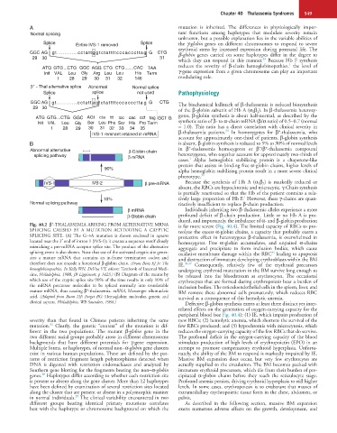
Fig. 40.2 β -THALASSEMIA ARISING FROM ALTERNATIVE MRNA is far more severe (Fig. 40.4). The limited capacity of RBCs to pro-
SPLICING CAUSED BY A MUTATION ACTIVATING A CRYPTIC teolyze the excess α-globin chains, a capacity that probably exerts a
SPLICING SITE. (A) The G→A mutation is shown enclosed in squares protective effect in heterozygous β-thalassemia, is overwhelmed in
located near the 3′ end of intron 1 (IVS-1); it creates a sequence motif closely homozygotes. Free α-globin accumulates, and unpaired α-chains
mimicking a pre-mRNA acceptor splice site. The product of the alternative aggregate and precipitate to form inclusion bodies, which cause
splicing event is also shown. Note that use of the activated cryptic site gener- oxidative membrane damage within the RBC leading to apoptosis
35
ates a mature mRNA that contains an in-frame termination codon and and destruction of immature developing erythroblasts within the BM
therefore does not encode a functional β-globin chain. (From Benz EJ Jr: The IE. 36,37 Consequently, relatively few of the erythroid precursors
hemoglobinopathies. In Kelly WN, DeVita VT, editors: Textbook of Internal Medi- undergoing erythroid maturation in the BM survive long enough to
cine, Philadelphia, 1988, JB Lippincott, p 1423.) (B) Diagram of the means by be released into the bloodstream as erythrocytes. The occasional
which use of the cryptic splice site 90% of the time results in only 10% of erythrocytes that are formed during erythropoiesis bear a burden of
the mRNA precursor molecules to be spliced normally into translatable inclusion bodies. The reticuloendothelial cells in the spleen, liver, and
+
mature mRNA, thus causing β -thalassemia. mRNA, Messenger ribonucleic BM remove these abnormal cells prematurely, which reduces RBC
acid. (Adapted from Bunn HF, Forget BG: Hemoglobin: molecular, genetic and survival as a consequence of this hemolytic anemia.
clinical aspects. Philadelphia, WB Saunders, 1986.) Defective β-globin synthesis exerts at least three distinct yet inter-
related effects on the generation of oxygen-carrying capacity for the
peripheral blood (see Fig. 40.4): (1) IE, which impairs production of
severity than that found in Chinese patients inheriting the same new RBCs; (2) hemolytic anemia, which shortens the survival of the
32
mutation. Clearly, the genetic “context” of the mutation is dif- few RBCs produced; and (3) hypochromia with microcytosis, which
ferent in the two populations. The mutant β-globin gene in the reduces the oxygen-carrying capacity of the few RBCs that do survive.
two different racial groups probably arose in different chromosome The profound deficit in the oxygen-carrying capacity of the blood
backgrounds that have different potentials for γ-gene expression. stimulates production of high levels of erythropoietin (EPO) in an
Multiple forms, or haplotypes, of normal non–α-globin gene clusters attempt to promote compensatory erythroid hyperplasia. Unfortu-
exist in various human populations. These are defined by the pat- nately, the ability of the BM to respond is markedly impaired by IE.
terns of restriction fragment length polymorphisms detected when Massive BM expansion does occur, but very few erythrocytes are
DNA is digested with restriction endonucleases and analyzed by actually supplied to the circulation. The BM becomes packed with
Southern gene blotting for the fragments bearing the non–α-globin immature erythroid precursors, which die from their burden of pre-
28
genes. Haplotypes differ according to whether each restriction site cipitated α-globin chains before they reach the reticulocyte stage.
is present or absent along the gene cluster. More than 12 haplotypes Profound anemia persists, driving erythroid hyperplasia to still higher
have been defined by examination of several restriction sites located levels. In some cases, erythropoiesis is so exuberant that masses of
along the cluster that are present or absent in a polymorphic manner extramedullary erythropoietic tissue form in the chest, abdomen, or
28
in normal individuals. The clinical variability encountered in two pelvis.
different groups bearing identical primary mutations correlates As described in the following section, massive BM expansion
best with the haplotype or chromosome background on which the exerts numerous adverse effects on the growth, development, and