
Page 667 - Hematology_ Basic Principles and Practice ( PDFDrive )
P. 667
Chapter 40 Thalassemia Syndromes 569
no longer persists after transformation of MDS to acute leukemia. A
This finding suggests that the Hb H–producing clone does not have Lepore–anti-Lepore Gγ Aγ (δβ)
a selective survival or growth advantage. Gγ Aγ δ β N C
The hematologic phenotype, as reflected by the amount of Hb H N C Lepore
present in blood, of ATMDS is much more severe than that of the
486
ATR-X syndrome. Some of this difference in severity may be C
because of the nature of the ATMDS mutations, some of which are N Gγ Aγ δ β
null mutations that are likely to be lethal when present in germline N βδ β C
DNA of ATR-X embryos. However, the difference in severity is also Gγ Aγ δ
observed in the case of mutations found in both syndromes that are B Anti-Lepore
identical or similar in expected functional consequences. This finding Kenya Gγ (γβ)
suggests that additional abnormalities in gene expression in ATMDS Gγ Aγ δ β N C
contribute to the severity of the deficit in α-globin gene expression N C Kenya
observed in this syndrome. Perhaps the responsible defective
cofactor(s) is one or more of the proteins that interact with the ATRX N C
protein to produce a fully functional macromolecular complex that Gγ Aγ δ β N C
can act as a transcriptional cofactor or that can influence the epigen- Gγ Aγ δ (βAγ) δ β
etic control of α-globin gene expression. “Anti-Kenya”
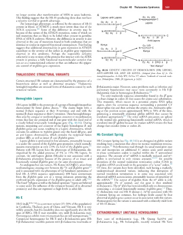
Fig. 40.16 GENETIC ORIGINS OF HEMOGLOBIN (HB) LEPORE,
THALASSEMIC STRUCTURAL VARIANTS ANTI-LEPORE HB, AND HB KENYA. (Adapted from Benz EJ Jr: The
hemoglobinopathies. In Kelly WN, DeVita VT, editors: Textbook of internal medi-
cine, Philadelphia, 1988, JB Lippincott, p 1423.)
Certain structural Hb variants are characterized by the presence of a
6
biosynthetic defect as well as abnormal structure. Thalassemic
hemoglobinopathies are unusual forms of thalassemia caused by such β-thalassemia major. However, some problems such as infection and
structural variants. pulmonary hypertension may occur more commonly in Hb E-β-
thalassemia than in homozygous β-thalassemia. 494
E
The only nucleotide sequence abnormality found in the β -gene
Hemoglobin Lepore is a base change in codon 26 that causes the amino acid substitution.
This mutation, which occurs in a potential cryptic RNA splice
Hb Lepore (α2βδ) is the prototype of a group of hemoglobinopathies region, alters the consensus sequence surrounding a potential GT
1–4
characterized by fused globin chains. The chains begin with a donor splice site and thus activates the cryptic site. Alternative splic-
normal δ-chain sequence at their N-terminus and end with the ing at this position occurs approximately 40% to 50% of the time,
normal β-chain sequence at their C-terminus. These hemoglobinopa- generating a structurally abnormal globin mRNA that cannot be
495
thies arise by unequal or nonhomologous crossover or recombination translated appropriately. The other mRNA precursors are spliced
events that fuse the proximal end of one gene with the distal end of at the normal site, generating functionally normal mRNA, which is
E
a closely linked structurally homologous gene (Fig. 40.16). During translated into β -globin because the mature mRNA retains the base
meiosis, mispairing and crossover of the highly homologous δ- and change that encodes lysine at codon 26.
β-globin genes can occur, resulting in a Lepore chromosome, which
contains (in addition to γ-globin genes) only the fused δβ gene, and
an anti-Lepore chromosome, which contains the reciprocal fusion Hb Constant Spring
product (δβ), as well as intact δ- and β-globin genes. 1
Lepore globin is synthesized in low amounts, presumably because Hb Constant Spring (see Fig. 40.14) is an elongated α-globin variant
it is under the control of the δ-globin gene promoter, which normally resulting from a mutation that alters the normal translation termina-
491
496
sustains transcription at only 2.5% the level of the β-globin gene. tion codon. Polyribosomes read through the usual translation stop
Patients with Hb Lepore have the phenotype of β-thalassemia, dis- site and incorporate an additional 31 amino acids until another
tinguished by the added presence of 5% to 15% Hb Lepore. In in-phase termination codon is reached within the 3′ untranslated
cs
cs
contrast, the anti-Lepore globin (Miyada) is not associated with a sequence. The amount of α mRNA is markedly reduced, and α -
β-thalassemia phenotype because of the presence of an intact and globin is synthesized in only minute amounts. 459,497 Six possible
functionally normal β-globin gene on the same chromosome. mutations of the normal translation termination codon (UAA) in
498
An analogous but rare variant, Hb Kenya [α2(Aγβ)2], arises from α-globin mRNA could result in the generation of a “sense” codon.
492
A
nonhomologous crossing over between the γ- and β-globin genes Of these, five variants have been identified, each having a markedly
and is associated with the phenotype of Gγ hereditary persistence of underproduced abnormal variant, indicating that disruption of
fetal Hb. A DNA sequence approximately 600 bases downstream normal translation termination is in some way associated with
from the β-globin gene acts as a strong enhancer, promoting the abnormal mRNA accumulation, presumably because of instability of
cs
459
erythroid-specific expression of the β-globin genes in adult cells. 3,4,21 the mRNA. The output of α-globin from the α allele is only
A
G
The fused γ β gene as well as the linked upstream γ gene are believed approximately 1% of normal, and the gene is thus rendered
cs
to come under the influence of the enhancer because of its abnormal α-thalassemic. The α allele has been identified only on chromosomes
1–4
proximity and thus are expressed at high levels in adult life. containing a cis-linked functionally normal α-globin gene. Thus,
+
cs
α -thalassemia trait and Hb H disease–(- /α α) associated with Hb
Constant Spring are common, but hydrops fetalis caused by four
Hb E abnormal α-globin genes cannot occur in association with this variant.
Homozygosity for the variant is associated with a relatively mild form
26Glu→Lys 1
Hb E (α 2β 2 ) is a common variant (15–30% of the population) of Hb H disease.
in Cambodia, Thailand, parts of China, and Vietnam. Hb E is very
mildly unstable, but this instability does not significantly alter the life
span of RBCs. Hb E trait resembles very mild β-thalassemia trait. EXTRAORDINARILY UNSTABLE HEMOGLOBINS
493
Homozygotes exhibit more microcytosis but are still asymptomatic.
Compound heterozygotes for Hb E and a β-thalassemia gene (Hb Rare cases of α-thalassemia (e.g., Hb Quong Sze)454 and
E-β-thalassemia) resemble patients with β-thalassemia intermedia or β-thalassemia (e.g., Hb Indianapolis, recently renamed Hb Terre