
Page 662 - Hematology_ Basic Principles and Practice ( PDFDrive )
P. 662
564 Part V Red Blood Cells
deferiprone, and deferasirox have all proved to be safe and effective β-Thalassemia Minor (Thalassemia Trait)
in thalassemia intermedia. 426–428
Thromboembolic events represent a major complication of thalas- Inheritance of a single β-thalassemia allele usually results in a mild
semia intermedia, occurring in 10% to 34% of patients. 326,429 These hypochromic microcytic anemia. The Hb level averages 1 or 2 g/dL
events include stroke, pulmonary embolism, portal vein thrombosis, lower than that seen in normal persons of the same age and gender.
and deep vein thrombosis of the legs. A hypercoagulable state may Hb F levels decline more slowly than usual in the first year of life,
also contribute to the pulmonary hypertension that commonly occurs and the diagnostic elevated Hb A 2 levels are established by approxi-
in patients with thalassemia intermedia and is the primary cause of mately 6 months of age. 443–445 Strong intrafamilial correlations of
430
congestive heart failure. Splenectomy is a risk factor for thrombo- both Hb A 2 and mean corpuscular volume (MCV) are noted. 446,447
embolic events in patients with thalassemia intermedia, resulting in Osmotic fragility is decreased; indeed, a one-tube osmotic fragility
444
thrombocytosis and allowing the prolonged circulation of damaged test has been used in the past for mass screening. The RBC count
326
RBCs that generate increased amounts of thrombin. Some inves- is increased or normal. The RBCs are characteristically hypochromic
tigators consider the risk of thromboembolic events after splenectomy (MCH <26 pg) and microcytic (MCV <75 fL). The smear shows
for thalassemia intermedia to be sufficiently high to warrant short- varying numbers of target cells, poikilocytes, ovalocytes, and baso-
term anticoagulation in the perioperative period and during preg- philic stippling (Fig. 40.13). The reticulocyte count is normal or
326
nancy. Oral contraceptives should be used with extreme caution, slightly elevated. RBC survival is normal, iron utilization is decreased,
445
if at all. Interestingly, known genetic thrombophilias in other popula- and slight IE is present. During pregnancy, the anemia of thalas-
tions like factor V Leiden, the prothrombin gene mutation 20210, semia trait often becomes more severe, but transfusions are rarely
and MTHFR C677T mutations have not been associated with necessary. Increased folic acid supplementation may improve Hb
thrombotic risk in this population. 431 during this period. Because iron deficiency may occur during preg-
Extension of hematopoietic tissue beyond the confines of the nancy, iron supplementation has been advised to avoid compounding
bones occurs in patients with thalassemia intermedia as a result of the causes of anemia. 448,449 In general, thalassemia trait carries no
the intense erythropoiesis. This complication occurs less frequently direct clinical symptoms or pathologic consequences for the patient.
in patients with thalassemia major because of the partial suppression Studies have suggested there may be an increased tendency for gall-
of erythropoiesis by regular transfusions. Masses of extramedullary stones and cholecystitis, but otherwise this condition should be
17
hematopoietic tissue develop in the spinal epidural space, thorax, largely asymptomatic. The diagnosis of thalassemia trait assumes
cranium, pelvis, and elsewhere. 367,432–442 These masses may be particular importance in women who are pregnant or considering
detected as incidental findings on imaging studies of the chest or pregnancy because of the potential for having a child with thalassemia
abdomen. 433–439 In other instances, the masses produce symptoms major.
by compressing neighboring structures. For example, patients with
extramedullary hematopoietic masses may develop paraplegia from
spinal cord compression or loss of visual acuity or visual fields caused α-THALASSEMIA SYNDROMES
by optic nerve compression. 432,435,441,442 Additional clinical presenta-
tions of hematopoietic masses include pleural effusions and upper The α-thalassemias are more difficult to diagnose because characteris-
airway obstruction. 437,438,440 Initiation of regular transfusions for tic elevations in Hb A 2 or Hb F, seen in many cases of β-thalassemia,
patients with thalassemia intermedia or intensification of the ongoing do not occur, making Hb electrophoresis difficult to use for
transfusion program for patients with thalassemia major reduces the diagnostic testing. However, the gene deletions responsible for the
size of extramedullary hematopoietic masses and helps to prevent most common varieties are readily detectable by molecular biology
recurrences. (Tables 40.4 and 40.5). methods. 450
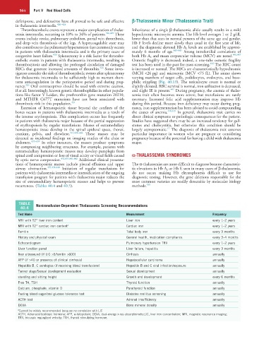
TABLE Nontransfusion-Dependent Thalassemia Screening Recommendations
40.4
Test Name Measurement Frequency
MRI with T2* liver iron content Liver iron every 1–2 years
MRI with T2* cardiac iron content a Cardiac iron every 1–2 years
Ferritin Total body iron every 3 months
History and physical exam General health, medication compliance every 3–4 months
Echocardiogram Pulmonary hypertension TRV every 1–2 years
Liver function panel Liver failure, hepatitis every 3 months
liver ultrasound (if LIC >5/ferritin >800) Cirrhosis annually
AFP (if >40 or presence of clinical cirrhosis) Hepatocellular carcinoma annually
Hepatitis B, C serologies (if receiving blood transfusions) Hepatitis B and C viral infection/exposure annually
Tanner stage/Sexual development evaluation Sexual development annually
standing and sitting height Growth and development every 6 months
Free T4, TSH Thyroid function annually
Calcium, phosphate, vitamin D Parathyroid function annually
Fasting blood sugar/oral glucose tolerance test Diabetes mellitus screening annually
ACTH test Adrenal insufficiency annually
DEXA Bone mineral density annually
a Cannot be widely recommended because no correlation with LIC
ACTH, Adrenocorticotropic hormone; AFP, α-fetoprotein; DEXA, dual-energy x-ray absorptiometry;LIC, liver iron concentration; MRI, magnetic resonance imaging;
TRV, tricuspic regurgitant velocity; TSH, thyroid stimulating hormone.