
Page 681 - Hematology_ Basic Principles and Practice ( PDFDrive )
P. 681
Chapter 41 Pathobiology of Sickle Cell Disease 573
Hemoglobin S Charge and Tetramer Assembly
Hemoglobin S Solubility and Hemoglobin S
Formation of Hb tetramers requires proximate assembly of stable Polymerization
dimers from unlike monomers (e.g., α + β → αβ), an event governed
by electrostatic attraction. The normal α and β chains are positively Oxy-HbS, oxy-HbA, and deoxy-HbA have very high solubilities, but
and negatively charged, respectively. In heterozygous states for deoxy-HbS aggregates into densely packed polymers, a process that is
3,4
β-globin mutants, β-chain competition for dimer assembly is a fully reversible with reoxygenation. This abnormal property causes
1
determinant of the relative proportions of the Hb variants. Mutant the eponymous RBC shape change from polymer-mediated distor-
β chains with lowered negative charge form αβ dimers more slowly; tion, the fundamental basis for disease promotion in sickling disorders.
C
A
A
S
the relative rates for dimer association are αβ >αβ >αβ , with αβ
S
dimers formed about twice as rapidly as αβ dimers. This explains
why those with sickle trait typically have only 40% HbS and why the Polymer Structure
proportion of HbS exceeds this in HbSC disease. It also explains the
effect of concurrent α-thalassemia on the proportion of HbS in sickle Deoxygenation transforms soluble HbS into a highly viscous and
trait; as availability of α chains becomes limiting, the percentage of semisolid gel that behaves thermodynamically similar to a crystal in
HbS typically drops from 40% to 35% (one α deletion), 30% (two equilibrium with a solution of individual tetrameric Hb molecules.
α deletions), or less than 25% (three α deletions). Even complete deoxygenation does not convert all deoxy-HbS to
polymer. The insoluble phase is a collection of domains of aligned
polymers, the basic unit of which is a double strand in which two
Hemoglobin S Stability and Oxidant Formation strings of deoxy-Hb tetramers make multiple contacts with each other
(Fig. 41.3).
S
HbS is modestly unstable, observed in vitro as instability to various Each HbS tetramer has two β chains, the β 1 and β 2 . Deoxy-HbS
applied stresses. Two stresses that are most clearly physiologic involve undergoes a slight structural shift so that the A helix β 6Val “donor”
2
Hb oxidation. HbS has an abnormal redox potential compared with site of the β 2 chain in one tetramer can contact an EF helix “acceptor”
HbA that may underlie its only modestly (~40%) increased auto- site (formed mainly by β 85Phe , β 88Leu , and β 70Ala ) in the β 1 chain of a
oxidation rate. Yet, HbS exhibits markedly (~340%) augmented tetramer in the neighboring single string. This critical, lateral associa-
instability and oxidation upon interaction with aminophospholipids tion can be made only when HbS is in its deoxy conformation; the
characteristic of the membrane’s inner leaflet. Its behavior once it EF helix hydrophobic pocket is not a favorable acceptor site for the
A
enters the plasma environment (caused by intravascular hemolysis) is charged β 6Glu of the β in HbA. In HbS, the β 6Val in the β 1 subunit
unknown. Although the physical–chemical mechanism of the desta- is located so it cannot participate in such contacts. However, the β 2
6
bilizing role of the β valine in HbS is not known, this instability chain of the second single string can form chemically similar β 6Val -
leads to accumulation of various Hb and iron forms at the cytosol– dependent contacts with the β 1 chain of the first single string. There
2
membrane interface. The resulting occurrence of abnormal, oxidative are multiple additional axial and lateral contacts, but these are largely
biochemistry promotes a number of prominent defects of the sickle the same for deoxy-HbA and deoxy-HbS and are not themselves
RBC membrane. sufficient to stabilize a polymeric structure.
b 1
a 1
b 2
a 2 b 1
a 1
b 2
b 1 a 2
a 1
b 2
a 2 b 1
a 1
b 2
b 1 a 2
a 1
b 2
a 2 b 1
a 1
b 2
a 2
=b 6 Val
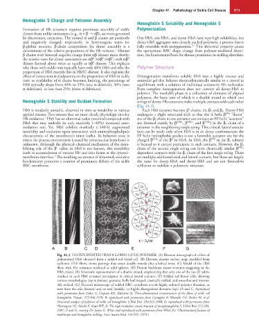
A B C D E F G
Fig. 41.3 DEOXYGENATED HEMOGLOBIN S (HbS) POLYMER. (A) Electron micrograph of a fiber of
polymerized HbS obtained from a sickled red blood cell. (B) Electron density surface map, modeled from
authentic HbS fibers, shows pairings that create double strands plus a helical twist. (C) Model of the HbS
fiber, with Hb tetramers rendered as solid spheres. (D) Protein backbone shows tetramer staggering in the
6
HbS crystal. (E) Schematic representation of a double strand, emphasizing that only one of the two β valine
residues in each HbS tetramer participates in critical lateral contacts. (F) Sickled red blood cells, showing
various morphologies (top to bottom): granular, holly leaf shaped, classically sickled, and smoother and irrevers-
ibly sickled. (G) Electron microscopy of sickled RBC cytoplasm reveals highly ordered polymer domains, as
seen from the side (bottom) and on end (middle), or highly disorganized domains (top). (A and C, Reproduced
with permission from Dykes G, Crepeau RH, Edelstein SJ: Three-dimensional reconstruction of the fibres of sickle cell
hemoglobin. Nature 272:506,1978; B, reproduced with permission from Carragher B, Bluemke DA, Becker M, et al:
Structural analysis of polymers of sickle cell hemoglobin. J Mol Biol 199:315,1988; D, reproduced with permission from
Harrington DJ, Adachi K, Royer WE, Jr: The high resolution crystal structure of deoxyhemoglobin S. J Mol Biol 272:398,
1997; F and G, courtesy Dr. James G. White and reproduced with permission from White JG: Ultrastructural features of
erythrocyte and hemoglobin sickling. Arch Intern Med 133:545, 1974.)