
Page 685 - Hematology_ Basic Principles and Practice ( PDFDrive )
P. 685
Chapter 41 Pathobiology of Sickle Cell Disease 577
Anti– Hemoglobin
band 3 precipitate
Bivalently
bound 30 µm
anti–
band 3
Band 3
cluster
Band 3
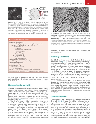
Fig. 41.7 BAND 3 AND IMMUNOGLOBULIN COCLUSTERING.
Denatured Hb on the RBC membrane, is associated with clumping of Band
3, and opsonization by naturally occurring anti-Band 3 antibody. Clusters of
band 3 are colocalized with immunoglobulin on the membranes of sickle red
blood cells (left). The drawing shows the colocalization scheme (right).
(Reproduced with permission from Schluter K, Drenckhahn D: Co-clustering of
denatured hemoglobin with band 3: Its role in binding of autoantibodies against band
3 to abnormal and aged erythrocytes. Proc Natl Acad Sci U S A 83:6137, 1986.) Fig. 41.8 RBC ADHESION TO ENDOTHELIUM. RBCs adhere to the
vascular wall endothelium under flow conditions in the microcirculation of
a rat infused with human cells. Immobile RBCs are on walls of the postcapil-
lary venule, and the smaller feeder microvessels (small arrows) have no flow
Major Sickle RBC Membrane Defects because of the logjam of RBC. (Reproduced with permission from Kaul DK, Fabry
ME, Nagel RL: Microvascular sites and characteristics of sickle cell adhesion to vascular
Membrane iron deposits → endothelium in shear flow conditions: Pathophysiological implications. Proc Natl Acad
Band 3 clumping → Ig attraction → erythrophagocytosis
Oxidative reactions targeted at membrane → Sci U S A 86:3356, 1989.)
Thiol oxidation →
ISC formation
↓ Deformability and ↑ fragility
PS externalization contributes to various sickling-induced RBC responses, e.g.,
Cation leak microvesiculation.
Microvesiculation
Lipid peroxidation →
Mechanosensitivity Irreversibly Sickled Cells
Erythrophagocytosis
Abnormal cation homeostasis → The sickled RBCs seen on a typically-obtained blood smear are
RBC dehydration → ↓ deformability mostly ISCs (see Fig. 41.1). Their permanent shape abnormality is
Abnormal microrheology → caused not by retained polymer but rather by membrane retention of
↓ Deformability
PS externalization → an elongated shape, explained by thiol oxidation of β-actin such that
Coagulation acceleration the spectrin–actin-4.1 complex exhibits abnormally slow dissocia-
Erythrophagocytosis tion. Otherwise, ISCs are similar to other equally dense RBC in
Adhesion to endothelium, monocytes, and macrophages having high MCHC, poor deformability, externalized PS, and low
Enhanced mechanosensitivity → ↑ responsiveness to deformation HbF content. ISC counts on average are higher in male patients,
perhaps reflecting their average lower levels of HbF. The fundamental
requirements for ISC formation seem to be RBC dehydration, pro-
longed deoxygenation, and assumption of a fixed membrane shape.
the bilayer from the underlying skeleton due to spicules of polymer- Perhaps there is a prior “conditioning” residence in the
ized hemoglobin, with enhanced susceptibility caused by protein microcirculation.
thiol oxidation. The clinical importance of ISCs lies in their ability to prompt
diagnosis of a sickling disorder when seen on blood smear and in
their short life span that contributes to overall hemolytic rate. They
Membrane Proteins and Lipids would contribute to the RBC logjam involved in occlusion, but it is
unclear whether ISC count correlates with vasoocclusive manifesta-
Sickle RBC membrane protein function is adversely affected by thiol tions. Although still adhesive to endothelium, ISCs are less so than
2
oxidation and possibly other oxidative protein modifications. other sickle subpopulations, but they exhibit greater adherence to
Ankyrin interactions with spectrin and Band 3 are abnormal, gly- macrophages.
cophorin and Band 3 exhibit decreased mobility, and thiol-oxidized
β-actin displays abnormal associations in the spectrin–actin-4.1
complex. Band 3 is abnormally clumped from binding of denatured Endothelial Adhesivity
HbS, which enables attraction of naturally occurring anti-Band 3
immunoglobulin (Fig. 41.7). Oxygenated sickle RBCs are abnormally adhesive to vascular endo-
Normal enforcement of bilayer phospholipid asymmetry is thelial cells (Fig. 41.8). About 20 candidate mechanisms have been
impaired in sickle RBCs. A scramblase that moves phosphatidylserine implicated, most involving adhesion molecules on endothelium,
(PS) outward is activated by calcium transits, and a translocase that adhesive structures restricted to reticulocytes or present on all RBCs,
8
restores PS inwardly can be inhibited by thiol oxidation. RBC sickling and with or without bridging by adhesogenic plasma proteins. Some
promotes PS externalization, especially in ISCs, but also in some mechanisms require RBC signaling responses to plasma factors for
reticulocytes. Other changes include presence of peroxidation activation. Involvement of mixed cell interactions with endothelium
byproducts such as malondialdehyde (MDA) that can cross-link has been proposed. Most described candidate mechanisms involve
proteins. Notably, the increased presence of bilayer lipid hydroperox- adhesive reticulocytes and are high affinity, identified using flowing
ides appears to account for the sickle RBC membrane’s abnormal conditions. Yet, in the biologic context microcirculatory blood flow
mechanosensitivity, evident in its enhanced cation leak response can be intermittent and occurs within vessels of constraining diam-
to deforming stress. Presumably, this deformation susceptibility eters enabling greater potential contact surface area. It seems probable