
Page 684 - Hematology_ Basic Principles and Practice ( PDFDrive )
P. 684
576 Part V Red Blood Cells
capacity. It is difficult to predict whether this produces a net benefit
or loss. RBC and Hb appear to participate in NO transport to
the microcirculation, although both magnitude of the effect and
mechanisms involved are debated. NO is asserted to improve RBC
deformability and impair HbS polymerization. Reaction of NO with
oxy-Hb causes Hb oxidation to met-Hb and reciprocal consumption
of NO. 6
ABNORMALITIES OF SICKLE RED BLOOD CELLS
Even oxygenated sickle RBCs exhibit a variety of cellular and mem-
brane abnormalities that contribute directly to pathophysiology. A B C D
Some are the consequence of proximate polymer formation, while
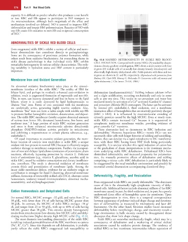
others result from oxidative biochemistry. An overarching theme in Fig. 41.6 MARKED HETEROGENEITY IN SICKLE RED BLOOD
sickle disease pathobiology is that individual sickle RBC exhibit CELL HYDRATION. Compared with normal RBCs (A) studied by discon-
remarkable heterogeneity in various cellular characteristics. The strik- tinuous density-gradient centrifugation, RBCs from a sickle patient with four
ing variability in hydration status and HbF content is particularly α genes (D) include cells of unusually low density (mostly reticulocytes) and
important. abnormally high density (dehydrated cells). Sickle patients with three and two
α genes are shown in (C and B), respectively. (Reproduced with permission from
Membrane Iron and Oxidant Generation Embury SH, Clark MR, Monroy G, Mohandas N: Concurrent sickle cell anemia and
alpha-thalassemia. J Clin Invest 73:116, 1984.)
An abnormal oxidative biochemistry takes place at the cytosol–
2
membrane interface of the sickle RBC. The avidity of HbS for
7
bilayer lipid, and perhaps its modestly enhanced auto-oxidation in deformation (mechanosensitivity). Sickling induces calcium influx
solution, result in augmented formation of superoxide and met-Hb. and a slight acidification, occurring stochastically and only in some
This, in turn, can become denatured and lose its heme to the lipid cells at any one time. This results in net potassium and water loss
2+
+
bilayer, where it is easily destroyed by lipid hydroperoxides to mediated mostly by activation of a Ca activated (Gardos) K channel
liberate “free” iron. Forms of iron associated with the membrane and potassium chloride (KCl) cotransport. The latter can be activated
are catalytically active, generating highly reactive oxidants. Also, by lowered pH, endothelin-1, thiol oxidation, and a membrane
membrane “free” iron can form a redox couple with soluble oxy-Hb interaction effect of hemoglobins that are relatively positively charged
to promote further hemoglobin oxidation, denaturation, and deposi- (HbC > HbS). It is influenced by macromolecular crowding of
tion. The sickle RBC membrane thereby acquires abnormal amounts cytosolic proteins caused by the high MCHC. Even at steady state,
2+
of various iron forms: Hb, denatured hemichrome, free heme, and sickle RBCs contain increased Ca because it is sequestered in
2
nonheme iron. A large portion of sickle RBC oxidant generation cytoplasmic inside-out membrane vesicles, providing evidence of
2+
and stress is from enhanced nicotinamide adenine dinucleotide prior cytosolic Ca transients.
phosphate (NADPH)-oxidase activity, probably in reticulocytes These aberrancies lead to decrements in RBC hydration and
7
and exhibiting a responsiveness to certain plasma substances, e.g., deformability. However, hyperdense RBCs—mostly ISCs—are not
endothelin-1. necessarily older cells with longer histories of sickling and unsickling.
Of equal importance to excessive oxidant generation, the mem- Rather, they can develop via a rapid reticulocyte-to-ISC transforma-
brane location of catalytic iron establishes in sickle RBC a unique tion, with those RBC having lower HbF levels being particularly
oxidant risk (not present in normal RBC) because it effectively targets susceptible. It is unclear whether this rapid induction of cation loss
oxidative damage to membrane components. Further, the juxtaposi- or the gradualism of classic interpretations is the dominant mecha-
tion of iron with bilayer lipid allows reinitiation of peroxidative chain nism underlying sickle RBC dehydration. Dehydrated RBCs have
reactions, effectively bypassing protection by vitamin E. Deficient diminished deformability and increased propensity for polymeriza-
levels of antioxidants (e.g., vitamin E, glutathione, ascorbic acid) in tion, the mutually promotive effects of dehydration and sickling
sickle RBC, caused by oxidative consumption and dietary insufficien- comprising a vicious cycle. RBC dehydration is particularly likely to
cies, contribute. The result is abnormal oxidation of membrane be exaggerated by the renal medullary environment and possibly by
protein thiols and peroxidation of membrane lipids. Among the nocturnal arterial desaturation accompanying disordered sleep.
many sickle membrane defects, evidence for an oxidative origin or
contribution is strongest for Band 3 clustering, abnormal membrane
stiffness, formation of irreversibly sickled cells (ISCs), aberrant cation Deformability, Fragility, and Vesiculation
homeostasis, tendency toward microvesiculation, abnormal mecha-
7
nosensitivity, and erythrophagocytosis. 2 Even oxygenated sickle RBCs are poorly deformable. The dominant
cause of this is the abnormally high cytoplasmic viscosity of dehy-
drated cells. Additional factors include abnormal stiffness of the RBC
Cation Homeostasis and Dehydrated Cells membrane caused, in part, by thiol oxidation and, in part, by a poorly
understood direct effect of hemoglobin upon the membrane. Upon
For normal RBCs, MCHC averages ~32 g/dL and varies from 27 to RBC deoxygenation in vitro, there is a temporal correspondence
38 g/dL, with fewer than 1% of cells having MCHC greater than between appearance of polymer-induced shape change and deteriora-
38 g/dL. In contrast, the MCHC of sickle RBCs averages ~34 g/ tion of deformability, as measured by micropipette and laser dif-
dL and ranges from 23 to 50 g/dL, with up to 40% of cells having fractometer. On the other hand, filtration studies found decreased
MCHC greater than 38 g/dL. This extreme density heterogeneity deformability before morphologic change, and viscometry reveals a
results from reticulocytosis (low-density, low-MCHC cells) and dehy- large deterioration in bulk viscosity caused by deoxygenated dense
drating mechanisms (higher density, high-MCHC cells) (Fig. 41.6). discocytes that show little shape change.
The most dramatic ion-handling abnormality of the sickle RBC Sickle RBCs are somewhat mechanically fragile, which may be a
is sickling-induced permeabilization of the RBC membrane to cations consequence of dehydration and a weakening of critical skeletal
+
+
2+
(Na , K , Ca ). Since this depends on cell deformation, it probably associations caused by oxidative protein damage. The tendency of
partly reflects the sickle RBC’s exaggerated leak susceptibility to sickled RBCs to lose membrane microvesicles reflects separation of