
Page 757 - Hematology_ Basic Principles and Practice ( PDFDrive )
P. 757
644 Part V Red Blood Cells
LCAT deficiency should be distinguished from an acquired deficiency preferentially intercalate into the inner half of the asymmetric lipid
of this enzyme, which is found in patients with severe liver disease. bilayer, expanding its surface area relative to that of the outer half of
the bilayer.
Stomatocytosis and Related Disorders
Hereditary Stomatocytosis-Hydrocytosis
Stomatocytes were first described in a girl with dominantly inherited
hemolytic anemia. On blood films, her RBCs contained a wide Hereditary hydrocytosis designates a heterogeneous group of heredi-
transverse slit or stoma (Fig. 45.9). In a three-dimensional view, these tary hemolytic anemias that are transmitted in an autosomal dominant
cells have a shape of a cup or a bowl. The slit-like appearance is an manner. The disorder is characterized by a moderate to severe hemo-
artifact that results from folding of the cells during blood smear lytic anemia with 10% to 30% stomatocytes (see Fig. 45.9), an elevated
preparation. MCV, and a reduced MCHC. Osmotic fragility of RBCs is markedly
Stomatocytes are seen in a variety of acquired and inherited dis- increased, as some of the swollen RBCs approach their critical
orders. The latter are often associated with abnormalities in RBC hemolytic volume. For unexplained reasons, RBC membrane lipids
cation permeability that lead to changes in RBC volume, which can and consequently membrane surface area are also increased, but this
be either increased (hence the designation hydrocytosis or overhy- increase in surface area is insufficient to correct the osmotic fragility
drated stomatocytosis) or decreased (xerocytosis or desiccytosis of the RBCs. RBC deformability is decreased.
[dessicate]), or in some cases near normal. The principal cellular lesion involves a marked increase in intracel-
There is no unifying theory to explain this morphologic abnor- lular sodium and water content with a mild decrease in intracellular
mality. In vitro, stomatocytes can be produced by drugs that potassium as a result of a marked sodium influx into the RBCs.
Despite a marked compensatory increase in active transport of
+
+
sodium (Na) and potassium by the Na /K -ATPase (which normally
maintains the low sodium and high potassium concentrations in the
cells) and an ensuing increase in glycolysis, the pump hyperactivity
is unable to compensate for the vastly increased sodium leak. Stoma-
tin (also known as band 7.2b), an integral membrane protein, is
decreased or absent from the erythrocyte membranes of most affected
patients. This deficiency appears to be a maturational loss in the bone
marrow and in the circulation, perhaps because of a defect in cellular
trafficking. Stomatin gene mutations have not been found in unre-
lated stomatocytosis patients deficient in this protein.
In some patients with hereditary hydrocytosis, missense mutations
in RhAG, I61R, or F65S, have been found. In oocytes these muta-
tions induce a monovalent cation leak, possibly opening the pore of
an ammonium transporter. Additional studies suggest that the F65S
mutation exhibits a gain-of-function phenotype with increased cation
conductance/permeability.
Splenectomy can improve, but not fully correct, the hemolysis. In
some patients, splenectomy can be deleterious or even contraindicated
(see later), perhaps because of altered endothelial cell adherence and
membrane phospholipid asymmetry.
Hereditary Xerocytosis and the
Intermediate Syndromes
Hereditary xerocytosis or desiccytosis describes an autosomal dominant
hemolytic anemia characterized by RBC dehydration and decreased
osmotic fragility. Affected individuals have characteristically moder-
ate to severe hemolysis with an increased MCHC, reflecting cellular
dehydration. Hydrops fetalis with fetal anemia or fetal ascites or the
presence of pseudohyperkalemia have been reported in a number
of xerocytosis kindred. Frequently the MCV is mildly increased.
In Coulter-type electronic counters, the conversion of pulse height
(from the resistance of a cell passing through an electric field) to a
cellular volume is dependent on cell shape. Xerocytes do not deform
to the same degree as normal cells, which causes the MCV to be
approximately 10% too high. The peripheral blood film (see Fig.
45.9) does not always reveal stomatocytes (which are more prominent
on wet films), but frequently target cells, dessicytosis (dessicate),
and spiculated cells are seen. In some of the cells, hemoglobin is
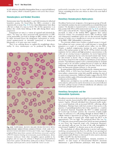
Fig. 45.9 PERIPHERAL BLOOD SMEARS FROM PATIENTS WITH concentrated (“puddled”) in discrete areas on the cell periphery.
HEREDITARY XEROCYTOSIS (DESICCYTOSIS) (TOP) AND STO- The mechanism of cellular dehydration is unclear and complex,
MATOCYTOSIS (HYDROCYTOSIS) (BOTTOM). (Top) A Wright-stained involving a net potassium loss from the cells that is not accompanied
peripheral blood smear from a patient with hereditary xerocytosis caused by by a proportional gain of sodium. Consequently, the net intracellular
a PIEZO1 mutation showing rare stomatocytes, occasional dessicytes—dense, cation content and cell water are decreased. In some reports a decrease
abnormal erythrocyte forms where hemoglobin appears puddled at the in RBC 2,3-DPG has also been noted.
periphery, and rare target cells. (Bottom) A Wright-stained peripheral blood Most HX patients have heterozygous missense mutations in
smear from a patient with hereditary hydrocytosis is shown. Numerous sto- PIEZO1. In vitro studies of HX-associated PIEZO1 mutations
matocytes, erythrocytes with a central mouth-like “stoma” are seen. demonstrate a gain-of-function phenotype, with many mutants