
Page 762 - Hematology_ Basic Principles and Practice ( PDFDrive )
P. 762
Chapter 46 Autoimmune Hemolytic Anemia 649
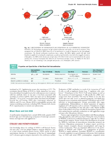
A
B C
Light coat Heavy coat
(no complement) (plus complement)
Splenic removal Hepatic removal
(slow circulation) (fast circulation)
Fig. 46.1 MECHANISM OF EXTRAVASCULAR HEMOLYSIS IN AUTOIMMUNE HEMOLYTIC
ANEMIA. (A) Macrophage encounters an IgG-coated erythrocyte and binds to it via its Fc receptors. Thus
entrapped, the red blood cell (RBC) loses bits of its membrane as a result of digestion by the macrophage’s
ectoenzymes. The discoid erythrocyte transforms into a sphere. (B) RBCs lightly coated with IgG (and
therefore incapable of activating the complement cascade) are preferentially removed in the sluggish circulation
of the spleen. (C) RBCs with a heavy coat of IgG; thus, C3b (black circles) can be removed both by the spleen
and the liver. (Courtesy Cunningham MJ, Silberstein LE: Autoimmune hemolytic anemia. In: Hoffman R, Benz EJ Jr,
Shattil SJ, et al, eds: Hematology: basic principles and practice, ed 4, Philadelphia, 2005, Elsevier.)
TABLE Properties and Specificities of Red Blood Cell Autoantibodies
46.1
Immunoglobulin Site of Removal of
(Subclass) Type of Antibody Clonality Specificity Hemolysis Red Blood Cells
WAIHAs IgG (1–4) , IgA Incomplete Mostly polyclonal Rh-antigens and Extravascular Spleen (liver)
non-Rh-antigens
CAIHAs IgM Complete Mostly clonal Anti-i, -I , -l, -Pr Intravascular Liver
T
Donath–Landsteiner antibody IgG — Polyclonal P antigen Intravascular —
CAIHA, Cold autoimmune hemolytic anemia; Ig, immunoglobulin; WAIHA, warm autoimmune hemolytic anemia.
incubated at 4°C. Agglutination occurs after warming to 37°C. The Production of RBC antibodies is a result of the interaction of T and
prominent clinical feature of PCH is a brisk, immediate but some- B cells, as well as regulatory factors (e.g., T regulatory cells, cyto-
17
times also delayed hemoglobinuria after cold exposure even in patients kines). Disturbances of the Th1/2 T-cell subset balance as well as
with low antibody titers. In the past, it has been associated with the occurrence of clonal regulatory T cells specific for a RBC auto-
secondary or tertiary syphilis. Now, two types can be distinguished antigen have been described. This may be linked to the fact that
15
clinically : (1) an acute, severe form (often associated with hemoglo- AIHA does not only occur in immunocompetent individuals but
binuria) but self-limiting AIHA after (respiratory) infections in frequently occurs in patients with acquired T-cell defects such as HIV
children, and (2) a rare, chronic AIHA in nonsyphilitic persons with infection or immunosuppressive therapy, particularly after organ
various underlying conditions, including NHL. Patients with chronic transplantation. Polymorphisms or altered expression of negative
PCH respond poorly to steroids and splenectomy. regulators of T-cell responses such as cytotoxic T lymphocyte antigen
4 (CTLA4) or interleukin-10 may also play a role. Mouse models
(New Zealand black mice) have revealed an association of genetic loci
Mixed Warm and Cold AIHA with antierythrocyte antibody production or cold agglutinin escape
tolerance after Mycoplasma infection.
A small number of patients have a mixed AIHA with a positive DAT Various target antigens have been described, with Rhesus polypep-
for both IgG and C3d indicating coexistence of warm IgG autoanti- tides, glycophorin, and erythrocyte band 3 being the most prominent
bodies and high-titer cold agglutinins. 16 in WAIHA. Cold reactive antibodies frequently target the I or i blood
group-specific antigens. Events linked to the development of second-
ary AIHA by induction of cross-tolerance (molecular mimicry) are
ETIOLOGY AND PATHOPHYSIOLOGY infections (Mycoplasma pneumoniae [I antigen target], parvovirus,
herpes viruses), neoplastic diseases (paraneoplasia), and drugs by
Our knowledge about the etiology of AIHA is still limited. Factors various mechanisms. There are important differences in the patho-
that may play a role are antigen mimicry; immune deficiency; and, genesis of WAIHA and CAIHA. The pathogenesis of primary
to a lesser extent, probably genetic factors. AIHA, similar to other WAIHA is largely unknown. Secondary WAIHA is a complication
autoimmune diseases, is a consequence of the loss of immunologic of several congenital or acquired immune deficiencies. Both moderate
(self-) tolerance against antigens expressed on the erythrocyte surface. (e.g., in CLL) and severe (HIV, posttransplant, congenital severe