
Page 753 - Hematology_ Basic Principles and Practice ( PDFDrive )
P. 753
640 Part V Red Blood Cells
the band 3 protein, inability to transport sulfate anions, and a mark- mutation is the first example of a defect of an integral membrane
edly restricted lateral and rotational mobility of band 3 protein in the protein leading to RBC membrane rigidity, an observation previously
membrane. attributed to properties of the membrane skeleton. The basis of the
increased rigidity is unclear.
The molecular basis of malaria resistance of SAO RBCs is likely
Laboratory Manifestations related to altered properties of the band 3 protein, which serves as
one of the malaria receptors, as evidenced by the inhibition of in vitro
The finding of 30% or greater of oval RBCs on the peripheral blood invasion by band 3-containing liposomes. In normal RBCs, the
film, some containing a central slit or a transverse ridge, in the context invasion process is associated with a marked membrane remodeling
of a notable absence of clinical and laboratory evidence of hemolysis that involves redistribution of intramembrane particles that contain
in a patient from the ethnic groups noted earlier is highly suggestive band 3 protein. Such particles cluster at the site of parasite invasion,
of the diagnosis. A useful screening test is the demonstration of the forming a ring around the orifice through which the parasite enters
resistance of ovalocytes or their ghosts to changes in shape produced the cell. The invaginated RBC membrane, which surrounds the
by treatments that produce spiculation in normal cells, such as meta- invading parasite, is free of intramembrane particles. The reduced
bolic depletion or exposure of ghosts to salt solutions. In contrast to lateral mobility of band 3 protein in SAO RBCs may preclude band
normal RBCs, which form spicules in response to such stimuli, SAO 3 receptor clustering, thereby preventing the attachment of the para-
RBCs or ghosts do not change shape after these treatments. The sites to the cells. Decreased exchange of anions across the RBC
mechanism of this resistance to changes in shape is not clear, and it membrane has also been proposed to contribute to the resistance of
may reflect the high rigidity of the RBC membrane. ovalocytes to malaria invasion. In addition, SAO RBCs consume ATP
Because the underlying cause of SAO is the deletion of 27 bases at a higher rate than normal cells, and the partial depletion of ATP
from the band 3 gene, isolation of genomic DNA or reticulocyte levels in ovalocytes has been suggested to account, at least in part, for
cDNA with subsequent amplification of the deletion-containing the resistance of these cells to malaria invasion in vitro.
region appears to be the most specific test for establishing the diag-
nosis of SAO. A single, severely affected homozygous SAO individual
who was transfusion dependent has been described. Acanthocytosis and Related Disorders
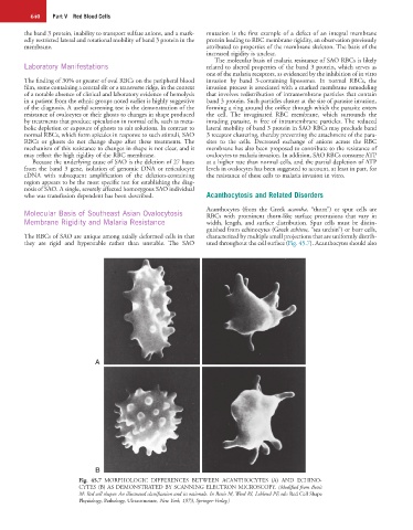
Molecular Basis of Southeast Asian Ovalocytosis Acanthocytes (from the Greek acantha, “thorn”) or spur cells are
RBCs with prominent thorn-like surface protrusions that vary in
Membrane Rigidity and Malaria Resistance width, length, and surface distribution. Spur cells must be distin-
guished from echinocytes (Greek echinos, “sea urchin”) or burr cells,
The RBCs of SAO are unique among axially deformed cells in that characterized by multiple small projections that are uniformly distrib-
they are rigid and hyperstable rather than unstable. The SAO uted throughout the cell surface (Fig. 45.7). Acanthocytes should also
A
B
Fig. 45.7 MORPHOLOGIC DIFFERENCES BETWEEN ACANTHOCYTES (A) AND ECHINO-
CYTES (B) AS DEMONSTRATED BY SCANNING ELECTRON MICROSCOPY. (Modified from Bessis
M: Red cell shapes: An illustrated classification and its rationale. In Bessis M, Weed RI, Leblond PF, eds: Red Cell Shape
Physiology, Pathology, Ultrastructure, New York, 1973, Springer-Verlag.)