
Page 754 - Hematology_ Basic Principles and Practice ( PDFDrive )
P. 754
Chapter 45 Red Blood Cell Membrane Disorders 641
be distinguished from keratocytes (“horn” red cells) that have few often markedly enlarged because of passive congestion as a result of
massive protuberances. underlying portal hypertension. Cholesterol also alters membrane
Acanthocytosis was first described in cases of abetalipoproteinemia permeability and interacts with several membrane skeletal proteins,
and subsequently in severe liver disease, the chorea-acanthocytosis but the role of these changes in spur cell lesions is unclear.
syndrome, the McLeod blood group phenotype, and other condi-
tions. The molecular mechanisms leading to acanthocytosis in
abetalipoproteinemia and severe liver disease have been extensively Clinical Manifestations
studied and have been attributed to changes in composition of
membrane lipids and their altered distribution between the two Most patients with chronic liver disease have a mild to moderate
hemileaflets of the lipid bilayer. anemia related to gastrointestinal blood loss, iron and folic acid
deficiencies, or hemodilution or as a direct effect of alcohol on RBC
precursors. Peripheral blood smears from these patients often reveal
Spur Cell Hemolytic Anemia of Severe Liver Disease target cells that are particularly prominent in obstructive jaundice.
In some patients, particularly those with end-stage liver disease,
Spur cell hemolytic anemia is an uncommon ominous complication anemia rapidly worsens and spur cells appear in high percentage in
of severe liver disease that is manifested by rapidly progressive hemo- the peripheral blood. This is accompanied by worsening jaundice,
lytic anemia and acanthocytes on the peripheral blood smear. rapid deterioration of liver function, hepatic encephalopathy, and
hemorrhagic diatheses. A similar clinical syndrome has been described
in patients with advanced metastatic liver disease, cardiac cirrhosis,
Pathobiology Wilson disease, fulminant hepatitis, and infantile cholestatic liver
disease. The development of spur cell hemolytic anemia is an ominous
The human RBC membrane contains nearly equal amounts of free sign in most patients, predicting a survival seldom exceeding weeks
(unesterified) cholesterol and phospholipids. The free cholesterol in to months. In theory, splenectomy could provide a marked improve-
the plasma readily equilibrates with the RBC membrane cholesterol ment, because the spleen is the major sequestration site of nonde-
pool. This is in contrast to esterified cholesterol, which cannot be formable acanthocytes; in reality, splenectomy is seldom considered
transferred from plasma into the RBC membrane. The plasma of because of severity of the underlying liver disease.
patients with severe liver disease contains abnormal lipoproteins that
have a high free cholesterol/phospholipid ratio. The excess free cho-
lesterol readily partitions into the RBC membrane, leading to a Abetalipoproteinemia
marked increase in free cholesterol in the cells. Consequently, normal
cells can develop a spur cell shape after their transfusion into a patient Bassen and Kornzweig first described an association of acanthocytosis
with severe liver disease or after incubation with the liver disease with atypical retinitis pigmentosa, progressive ataxic neurologic
patient’s plasma or cholesterol-enriched liposomes. disease, and a “celiac disease” later attributed to fat malabsorption.
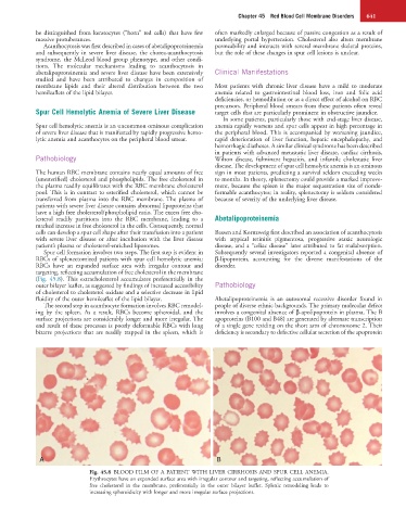
Spur cell formation involves two steps. The first step is evident in Subsequently several investigators reported a congenital absence of
RBCs of splenectomized patients with spur cell hemolytic anemia: β-lipoprotein, accounting for the diverse manifestations of the
RBCs have an expanded surface area with irregular contour and disorder.
targeting, reflecting accumulation of free cholesterol in the membrane
(Fig. 45.8). This extracholesterol accumulates preferentially in the
outer bilayer leaflet, as suggested by findings of increased accessibility Pathobiology
of cholesterol to cholesterol oxidase and a selective decrease in lipid
fluidity of the outer hemileaflet of the lipid bilayer. Abetalipoproteinemia is an autosomal recessive disorder found in
The second step in acanthocyte formation involves RBC remodel- people of diverse ethnic backgrounds. The primary molecular defect
ing by the spleen. As a result, RBCs become spheroidal, and the involves a congenital absence of β-apolipoprotein in plasma. The B
surface projections are considerably longer and more irregular. The apoproteins (B100 and B48) are generated by alternate transcription
end result of these processes is poorly deformable RBCs with long of a single gene residing on the short arm of chromosome 2. Their
bizarre projections that are readily trapped in the spleen, which is deficiency is secondary to defective cellular secretion of the apoprotein
A B
Fig. 45.8 BLOOD FILM OF A PATIENT WITH LIVER CIRRHOSIS AND SPUR CELL ANEMIA.
Erythrocytes have an expanded surface area with irregular contour and targeting, reflecting accumulation of
free cholesterol in the membrane, preferentially in the outer bilayer leaflet. Splenic remodeling leads to
increasing spheroidicity with longer and more irregular surface projections.