
Page 778 - Hematology_ Basic Principles and Practice ( PDFDrive )
P. 778
664 Part V Red Blood Cells
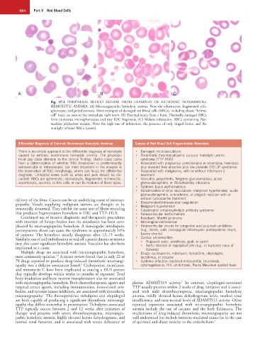
A B C
Fig. 47.1 PERIPHERAL BLOOD SMEARS FROM EXAMPLES OF EXTRINSIC NONIMMUNE
HEMOLYTIC ANEMIA. (A) Microangiopathic hemolytic anemia. Note the schistocytes, fragmented cells,
spherocyte, and polychromasia. More examples of damaged red blood cells (RBCs), including classic “helmet
cell” (top), are seen to the immediate right insert. (B) Thermal injury from a burn. Thermally damaged RBCs
form numerous microspherocytes and tiny RBC fragments. (C) Malaria infestation. RBCs containing Plas-
modium falciparum malaria. Note the high rate of infestation, the presence of only ringed forms, and the
multiply infested RBCs (center).
Differential Diagnosis of Extrinsic Nonimmune Hemolytic Anemias Causes of Red Blood Cell Fragmentation Hemolysis
There is no simple approach to the differential diagnosis of hemolysis • Damaged microvasculature
caused by extrinsic nonimmune hemolytic anemia. The physician • Thrombotic thrombocytopenic purpura–hemolytic uremic
must pay close attention to the clinical finding. Useful clues come syndrome (TTP–HUS)
from a determination of whether RBC breakdown is predominantly • Associated with pregnancy: preeclampsia or eclampsia; hemolysis
extravascular or intravascular, but most important in the analysis is plus elevated liver enzymes plus low platelets (HELLP syndrome)
the observation of RBC morphology, which can focus the differential • Associated with malignancy, with or without mitomycin C
diagnosis. Unhelpful terms such as aniso and poik should be dis- treatment
carded. RBCs are spherocytic, stomatocytic, fragmented, echinocytic, • Vasculitis: polyarteritis, Wegener granulomatosis, acute
acanthocytic, spurred, or bite cells, or can be mixtures of these types. glomerulonephritis, or Rickettsia-like infections
• Systemic lupus erythematosus
• Abnormalities of renal vasculature: malignant hypertension, acute
glomerulonephritis, scleroderma, or allograft rejection with or
delivery of the fetus. Cancer can be an underlying cause of microan- without cyclosporine treatment
giopathy. Vessels supplying malignant tumors are thought to be • Disseminated intravascular coagulation
• Malignant hypertension
structurally abnormal. They exhibit the same sort of fibrin stranding • Catastrophic antiphospholipid antibody syndrome
that produces fragmentation hemolysis in DIC and TTP–HUS. • Atrioventricular malformations
Continued use of invasive diagnostic and therapeutic procedures • Kasabach–Merritt syndrome
with insertion of foreign bodies into the circulation has been com- • Hemangioendotheliomas
plicated by microangiopathic hemolysis. A transjugular intrahepatic • Atrioventricular shunts for congenital and acquired conditions
portosystemic shunt can cause the syndrome in approximately 10% (e.g., stents, coils, transjugular intrahepatic portosystemic shunt,
of patients. The hemolysis usually disappears after 12–15 weeks. Levine shunts)
Similarly, use of coil embolization to seal off a patent ductus arteriosus • Cardiac abnormalities
may also cause significant hemolytic anemia. Vasculitis has also been • Replaced valve, prosthesis, graft, or patch
• Aortic stenosis or regurgitant jets (e.g., in ruptured sinus of
implicated as a cause. Valsalva)
Multiple drugs are associated with microangiopathic hemolysis, • Drugs: cyclosporine, mitomycin, ticlopidine, clopidogrel,
3,4
most commonly quinine. A recent review found that in only 22 of tacrolimus, or cocaine
78 drugs reported to produce drug-induced thrombotic microangi- • Systemic infection: bacterial endocarditis, brucellosis,
4
opathy was a definite association found. Cyclosporine, tacrolimus, cytomegalovirus, HIV, ehrlichiosis, Rocky Mountain spotted fever.
and mitomycin C have been implicated as causing a HUS picture
that typically develops within weeks to months of exposure. Total
body irradiation and bone marrow transplantation also are associated
6
with microangiopathic hemolysis. Both chemotherapeutic agents and plasma ADAMTS13 activity. In contrast, clopidogrel-associated
targeted cancer agents, including immunotoxins, monoclonal anti- TTP usually presents within 2 weeks of drug initiation and is associ-
bodies, and tyrosine kinase inhibitors, are associated with thrombotic ated with mild thrombocytopenia, microangiopathic hemolytic
5
microangiopathy. The thienopyridines ticlodipine and clopidogrel anemia, mildly elevated lactate dehydrogenase levels, marked renal
are both capable of producing a significant thrombotic microangi- insufficiency, and near-normal levels of ADAMTS13 activity. Other
opathy that differs somewhat in presentation. Ticlodipine-associated reported exposures associated with microangiopathic hemolytic
TTP typically occurs between 2 and 12 weeks after initiation of anemia include the use of cocaine and the herb Echinacea, The
therapy and presents with severe thrombocytopenia, microangio- mechanisms of drug-induced thrombotic microangiopathy are not
pathic hemolytic anemia, highly elevated lactate dehydrogenase, and well understood but include immune-mediated causes (as in the case
normal renal function, and is associated with severe deficiency of of quinine) and direct toxicity to the endothelium. 4