
Page 780 - Hematology_ Basic Principles and Practice ( PDFDrive )
P. 780
666 Part V Red Blood Cells
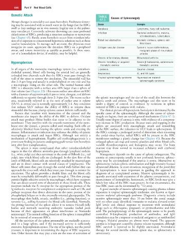
Osmotic Attack TABLE
47.1 Causes of Splenomegaly
Abrupt changes in osmolality can cause hemolysis. Freshwater drown-
ing may be associated with so much water in the lungs that the RBCs Cause Example
swell as they undergo an in vivo osmotic fragility test in the pulmo- Neoplasia Lymphoma, hairy cell leukemia
nary vasculature. Conversely, saltwater drowning can cause profound
dehydration of RBCs, producing a situation analogous to xerocytosis Infection Bacterial endocarditis, malaria,
(see Chapter 45). Rarely, acute hemolysis occurs from mistaken infu- schistosomiasis, tuberculosis
sion of or exposure to concentrated hypertonic solutions such as those Portal bed obstruction Alcoholic cirrhosis, splenic vein
used in hemodialysis. To manage such an event, the physician must thrombosis
recognize its cause, appreciate the shrunken RBCs on a peripheral Collagen vascular disease Systemic lupus erythematosus,
smear, and restore isotonicity as quickly as possible. In these cases, malignant phase of rheumatoid
use of a hemodialysis device, if available, may be helpful. arthritis
Chronic inflammatory disease Rheumatoid arthritis
Hypersplenism Chronic hereditary or acquired Severe β-thalassemia, autoimmune
hemolytic anemia hemolytic anemia
In all organs of the monocyte–macrophage system (i.e., reticuloen- Lipoidosis Gaucher disease
dothelial system), blood cells leaving the arterial bed are generally Amyloidosis AL and AA types
unloaded into channels such that the RBCs must pass through the
wall of the sinus to reenter the circulation. The sinusoidal wall has Tropical splenomegaly syndrome Hyperreactive malarial
slits 2–3 µm long and usually is endothelialized on one side and has splenomegaly syndrome
a macrophagic lining on the other side. The normal human adult
RBC is a discocyte with a surface area 40% larger than a sphere of
that volume (see Chapter 33). This excess surface area allows an RBC
with a diameter of approximately 8 µm to twist, elongate, and deform
sufficiently to squeeze through these 2–3-µm slits. The excess surface the splenic macrophages and the size of the small slits between the
area, occasionally referred to as the ratio of surface area to volume splenic cords and sinuses. The macrophages and slits seem to be
(SA:V), is critical and is normally approximately 1.4. Any condition under a degree of control, as evidenced by variations in splenic
that reduces SA:V reduces the ability of RBCs to traverse these removal of RBCs in patients with malaria.
sinusoidal slits because plump spheres cannot deform sufficiently. The clinical picture of hypersplenic hemolysis is dominated by the
Factors that interfere with interaction of the cytosol and the specific cause of the splenomegaly. Although the causes of spleno-
membrane also impair the ability of the RBC to deform. Oxidant megaly are legion, there are several general mechanisms (Table 47.1).
attack may produce Heinz bodies that come to lie adjacent to the Usually some degree of anemia is seen, with evidence of a compensa-
membrane. They interfere with the smooth movement of the mem- tory increase in RBC production. Because stasis and trapping in the
brane over the cytosol, a process called tank treading. Such cells are spleen are associated with macrophagic attack and remodeling
selectively blocked from leaving the splenic cords and entering the of the RBC surface, the reduction in SA:V leads to spherocytosis. If
sinuses. Inflammation or infection may enhance the ability of splenic the RBCs undergo a prolonged period of distortion when traversing
macrophages to attack and ingest RBCs. Although not strictly a the cordal–sinus barrier, tailed RBCs will be present as the RBC
mechanism of hypersplenism, Kupffer cell erythrophagocytosis is a membranes undergo a plastic change (see Chapter 45). Because the
prominent finding in patients undergoing graft-versus-host hemolysis enlarged spleen can trap and remove platelets and white blood cells,
seen after liver transplantation. variable thrombocytopenia and leukopenia may occur. The bone
The spleen is more complicated than other reticuloendothelial marrow may show normal to increased cellularity with erythroid
organs in that the afferent arterioles pass through lymphoid nodules hyperplasia.
(i.e., white pulp) and then terminate in the cords of Billroth (i.e., red Management depends on the cause of splenic enlargement. The
pulp), into which blood cells are discharged. In the slow flow of the anemia or pancytopenia usually is not profound; however, splenec-
cords of Billroth, blood cells are selectively attacked by macrophages tomy may be contemplated if the anemia is severe. Alternatives to
and are in direct contact with several classes of lymphocytes. The splenectomy include splenic embolization and high-intensity focused
blood cells must then pass through the cordal walls before they can ultrasound ablation. In most situations, recognition of the possibility
approach the sinus wall, which they must pass through to reenter the of hypersplenism is most important in guiding the approach to
circulation. The spleen provides a double filter, and the blood cells diagnosis of an unexplained anemia. Massive splenomegaly is fre-
must be remarkably deformable to pass through it. This slow passage quently associated with expansion of the plasma compartment, and
permits highly selective action by macrophages, which have receptors measurement of hemoglobin, hematocrit, or RBC levels may give a
that can detect several sorts of alterations in these blood cells. These falsely low value of the RBC mass present. In that circumstance, the
51
receptors include the Fc receptor for the appropriate portion of the true RBC mass can be determined by Cr assay.
Ig molecule, receptors for complement components such as C3b, and A good example of massive splenomegaly causing plasma volume
perhaps receptors that detect alterations in the outer portion of the expansion is tropical splenomegaly syndrome, also known as hyper-
phospholipid bilayer or in the externally oriented glycopeptides. The reactive malarial splenomegaly syndrome. Diagnostic criteria include
macrophage then holds, retards, modifies (i.e., pitting function), or massive splenomegaly more than 10 cm below the costal margin
removes (i.e., culling function) the blood cells identified. Normally, with no other cause identified; immunity to malaria; elevated serum
the pitting function of the spleen allows it to remove Howell–Jolly IgM levels; and clinical response to treatment with antimalarial
bodies and normally occurring endocytic vacuoles (called pocks drugs such as chloroquine, proguanil, or pyrimethamine and folic
because of their appearance on phase interference or Nomarski acid. The pathophysiology of the splenomegaly seems to be poorly
microscopy). The normal culling function of the spleen is exemplified controlled B-lymphocytic production of antibodies, and IgM
by its removal of senescent RBCs. stimulation may be a response to malarial antigens or an unidentified
All the activities of the spleen presumably are markedly accentu- mitogen. Malarial parasites are almost never found. The apparent
ated in a large spleen, and if the increased activity is sufficiently anemia is in large part caused by plasma volume expansion, although
extensive, hypersplenism ensues. The size of the spleen, not the portal RBC survival is reported to be slightly attenuated. Antimalarial
pressure, is important in determining the degree of RBC sequestra- therapy for several months reduces spleen size, so splenectomy is
tion. Other factors that may play a role are the state of activation of unnecessary.