
Page 991 - Hematology_ Basic Principles and Practice ( PDFDrive )
P. 991
874 Part VII Hematologic Malignancies
increased SMAD1 mRNA, in DLBCL cells obtained from patients. HATs and HDACs are two classes of enzymes that mediate the
SMAD1 is the major downstream effector of TGF-β. TGF-β exerts an acetylation and deacetylation, respectively, at evolutionarily-conserved
antiproliferative effect in most NHL, and loss of antiproliferative N-terminal lysine residues. Acetylated histones are negatively charged
response to TGF-β has been demonstrated in the majority of NHLs. and do not bind as tightly to negatively charged DNA, thereby
SMAD1 hypomethylation has been associated with an abundance of facilitating gene transcription. In contrast, deacetylated histones bind
SMAD1 mRNA and restoration of chemosensitivity in DLBCL cells closely to DNA, preventing transcription. Acetylation status of
after azacitidine “priming.” chromatin and hence gene transcription is dictated by balanced activ-
ity of HATs and HDACs. Acetylation of core histone bases has also
been implicated in chromatin assembly, DNA repair, and replication
Histone Deacetylase Inhibitors timing of specific genomic regions. Cross-talk also exists between
acetylation and ubiquitination. Thus, HDACs can decrease the half-
Histone acetyl transferases (HATs) control gene expression through the lives of substrates by exposing the lysine residue for ubiquitination.
modification of chromatin structure through acetylation of chromatin- Other crucial functions affected by the delicate balance between
bound histones. Regulation of acetylation is through histone deacety- HATs and HDACs include activation of the apoptotic program via
lation. Without the ability to fine tune histone binding to chromatin interaction between Ku70 and Bax; protein localization (nuclear vs.
regions, gene expression is perturbed. Inhibition of this process by a cytoplasm); and DNA binding of transcription factors such as p53,
group of agents termed histone deacetylation inhibitors alters gene E2F1, GATA1, RelA, YY1, and hormone receptors. There is a
expression in both normal and malignant cells. Often the result is growing list of nonhistone proteins that are modulated by HATs or
differentiation of malignant cells or induction of apoptosis. Given the HDACs. These include hypoxia-inducible factor-1α (HIF-1α),
long history of the use of differentiating agents in leukemias, for APL β-catenin, α-tubulin, Ku70, importin-α 7, cortactin, and, most
(retinoids), low-dose ara-C and azacytidine, it is not surprising that recently, HSP90. HATs and HDACs can be classified into subfamilies
newer HDIs have undergone extensive evaluation in leukemias. according to the presence of highly conserved structural motifs. Please
refer to Table 57.5 for details.
Posttranslational Histone and Nonhistone Protein
Modifications and Gene Transcription Aberrant Histone Acetyl Transferase and Histone
Deacetylase Activity in Hematologic Malignancies
Nucleosomes are regularly repeating, structural units of chromatin,
which are essential in packaging eukaryotic DNA. Each unit is Aberrant activity of HATs and HDACs resulting in aberrant gene
composed of 146 base pairs of DNA tightly wrapped around a core transcription is a hallmark of many cancers, including many hema-
histone octamer. Each histone octamer consists of two units each of tologic malignancies. Several chromosomal translocations in leukemia
histones H2A, H2B, H3, and H4, and each nucleosome in turn is that produce chimeric fusion oncoproteins have been shown to
connected to its neighbor by a short segment of linker DNA approxi- recruit HDACs to promoters and repress genes involved in cell cycle
mately 10–80 base pairs in length. Histone H1 binds and stabilizes growth inhibition and differentiation. For example, PML-RARα in
linker DNA. Each core histone has an N-terminal tail, which is lysine APL and AML1-ETO generated by t(8;21) translocation in AML
rich and positively charged. Specific amino acid residues at the recruit HDACs to their target genes, resulting in chromatin modifica-
N-terminal undergo a variety of enzymatic posttranslational modifi- tion and repression of genes, leading to blocked differentiation and
cations. Modifications can also occur within the globular domain of inhibition of apoptosis. HDACs have also been found in complexes
histones that make extensive contacts with DNA. Histone code is the with proteins that regulate cell cycle checkpoints such as Rb and its
name given to the combination of biochemical modifications affect- family members. Resistance to chemotherapy can occur because of
ing different histone residues that specify chromatin function. increased levels of thioredoxin, a thiol reductase, and decreased levels
However, it has been suggested that various postsynthesis histone of thioredoxin-binding protein (TBP-2) in many cancers; HDIs can
modifications be considered an epigenomic alphabet. Each modifica- reverse this phenomenon. These effects create a strong rationale for
tion is a letter, and the combination of modifications at a specified developing inhibitors of HDAC activity that would correct transcrip-
genomic region is a word that may have different functional meanings tional deregulation of genes involved in cell cycle regulation and
depending on the context. apoptosis as cancer therapeutic agents.
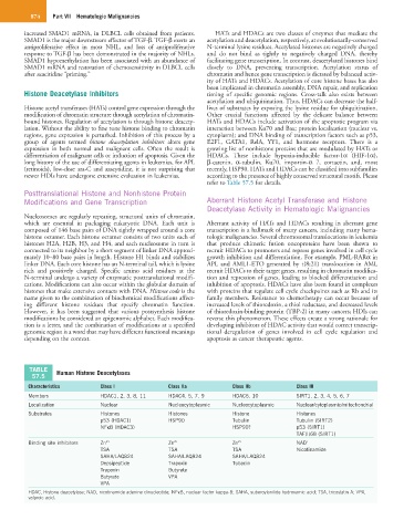
TABLE Human Histone Deacetylases
57.5
Characteristics Class I Class IIa Class IIb Class III
Members HDAC1, 2, 3, 8, 11 HDAC4, 5, 7, 9 HDAC6, 10 SIRT1, 2, 3, 4, 5, 6, 7
Localization Nuclear Nucleocytoplasmic Nucleocytoplasmic Nuclear/cytoplasmic/mitochondrial
Substrates Histones Histones Histone Histones
p53 (HDAC1) HSP90 Tubulin Tubulin (SIRT2)
NFκB (HDAC3) HSP90? p53 (SIRT1)
TAF(I)68 (SIRT1)
Binding site inhibitors Zn 2+ Zn 2+ Zn 2+ NAD +
TSA TSA TSA Nicotinamide
SAHA/LAQ824 SAHA/LAQ824 SAHA/LAQ824
Depsipeptide Trapoxin Tubacin
Trapoxin Butyrate
Butyrate VPA
VPA
HDAC, Histone deacetylase; NAD, nicotinamide adenine dinucleotide; NFκB, nuclear factor kappa-B; SAHA, suberoylanilide hydroxamic acid; TSA, tricostatin A; VPA,
valproic acid.