
Page 1018 - Williams Hematology ( PDFDrive )
P. 1018
992 Part VII: Neutrophils, Eosinophils, Basophils, and Mast Cells Chapter 65: Neutropenia and Neutrophilia 993
rare cases of biallelic mutations within the extracellular domain of the
G-CSF receptor which lead to nonresponse to treatment with G-CSF. 36
G-CSF is a very effective therapy for all of the recognized subtypes
of severe congenital neutropenia, increasing the neutrophil counts and
37
reducing recurrent fevers and infections. G-CSF acts to increase the
neutrophil counts by enhancing expression of a critical transcription
factor for granulopoiesis, C/EBPβ (CCAAT/enhancer binding protein
β), and the “emergency” pathway of myelopoiesis (as during steady state
38
C/EBPα is not functional). Approximately 5 percent of patients do
not respond to G-CSF. Hematopoietic transplantation is the only other
therapy known to improve the clinical course for these patients. 39,40
Untreated patients and patients treated with G-CSF are at risk for
developing acute myelogenous leukemia. The risk increases with time
41
on treatment with G-CSF, particularly in poorly responsive patients.
A novel molecular pathway of leukemogenesis was recently identified:
mutations in the hematopoietic cytokine receptor (G-CSFR) in com-
bination with the second mutations in the downstream hematopoietic
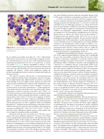
Figure 65–2. Morphology of a marrow sample from a patient with transcription factor (RUNX1), which could be used as a marker for
congenital neutropenia showing the maturation arrest at the level of identifying severe congenital neutropenia patients with a high risk of
promyelocytes. progressing to leukemia or myelodysplastic syndrome. 42
Congenital Immunodeficiency Diseases Neutropenia is a fea-
ture of the congenital immunodeficiency diseases and a contributing
the neutrophil count usually is less than 0.2 × 10 /L. Monocytosis, factor to their susceptibility to infections (Chap. 80). In most of these
17
9
mild anemia, thrombocytosis, and splenomegaly frequently are pres- conditions, neutropenia is attributed to a production disorder based
ent. Characteristically, the marrow shows early neutrophil precursors largely on histologic examination of the marrow. In X-linked agamma-
(myeloblasts, promyelocytes) but few or no myelocytes or mature globulinemia, which is attributed to defective B-cell development and
neutrophils (Fig. 65–2). Marrow eosinophilia is common. In vitro a mutation in a cytoplasmic (Bruton) tyrosine kinase (BTK), severe
43
marrow culture studies show poor growth in response to various growth neutropenia is present in approximately 25 percent of patients. Chil-
factors and with reduced numbers of marrow neutrophil and monocyte dren with common variable immunodeficiency often have neutropenia
18
43
progenitor cell colonies. Usually blood lymphocyte numbers are associated with thrombocytopenia and hemolytic anemia. Neutro-
normal, immunoglobulin levels are normal or increased, and lymphocyte penia occurs in almost half of patients with the X-linked hyperimmu-
functions are intact. noglobulin-M syndrome, a disorder caused by a mutation in the gene
The majority of patients with sporadic or autosomal dominant encoding the CD40 ligand. In severe combined immunodeficiency,
44
severe congenital neutropenia have heterozygous mutations of the gene neutropenia is not always present. The neutropenia varies over time
for neutrophil elastase (also called ELANE). Its product is a protease in individual patients. Neutropenia is particularly prominent in the
found normally in the neutrophil’s primary granules and lead to the rare immunodeficiency state, reticular dysgenesis. Neutropenia is a
43
induction of the unfolded protein response in the endoplasmatic retic- less-common feature of adenosine deaminase deficiency, the T−B+,
ulum. 15,19,20 A variety of mutations in exons 2 through 5 as well as in T−B−, Wiskott-Aldrich, and Omenn syndromes. 43,45,46 Neutropenia also
introns III and IV are the cause of this disease. 20–23 In the original Kost- occurs on an autoimmune basis in some cases of the Wiskott-Aldrich
mann family, and some other families with autosomal recessive disease, syndrome. 34,46 Mutations in the genes for growth factor independent
24
neutropenia is caused by mutations in the HAX-1 gene. HAX-1 is a protein-1 (GFI 1) can also cause neutropenia. 47
mitochondrial protein, and the mutations lead to accelerated apoptosis G-CSF therapy is effective in most patients with neutropenia asso-
of myeloid cells, as well as neurologic abnormalities. In addition, muta- ciated with these immunodeficiency syndromes.
tions in HAX-1 lead to defective G-CSF receptor signaling via HCLS1 Cartilage Hair Hypoplasia Syndrome This rare autosomal reces-
25
and LEF-1. Mutations in the gene for glucose-6-phosphatase catalytic sive disorder is characterized by short-limbed dwarfism, hyperexten-
subunit 3 (G6PC3) also cause severe neutropenia as a result of apoptosis sible digits, very fine hair, neutropenia, lymphopenia, and recurrent
of neutrophil precursors, as well as congenital cardiac and urogenital infections. The genetic locus is at 9p13 and affects a gene coding for
43
26
abnormalities. Additional autosomal-dominant, autosomal-recessive, an endoribonuclease. The degree of neutropenia is variable, with blood
9
X-linked, and sporadic forms have been described, with mutations in counts ranging from 0.1 to 2.0 × 10 /L. An accompanying defect in
27
31
30
other genes, including GFI1, WAS, p14, TAZ, JAGN1, TCIRG1, T-cell proliferation results from an abnormality in the transition from
32
28
29
and many others, although the genetic causes in many patients with the G to the G phase of the mitotic cycle. Patients have frequent bacte-
0
1
severe congenital neutropenia remain unidentified. rial and viral respiratory infections. Hematopoietic stem cell transplan-
Mutations in the gene for the receptor for G-CSF also occur in tation can correct the neutropenia and immune deficiency. 48,49
patients with severe congenital neutropenia 33,34 ; however, most of these Shwachman-Diamond Syndrome This autosomal recessive dis-
receptor mutations have caused truncations of the distal portion of the order combines short stature, pancreatic exocrine deficiency, and mar-
cytoplasmic domain of the receptor, an abnormality associated with row failure with neutropenia beginning early in the neonatal period. 50–53
altered sensitivity to G-CSF. G-CSF receptor mutations are part of the Thrombocytopenia and anemia may be severe (Chap. 35). The chromo-
evolution to myelodysplasia or acute myelogenous leukemia and are not somal locus of the mutation is at 7qll, and the mutation affects the SBDS
50
the primary cause of this neutropenia. An exception may be a patient gene. The mutation causes a proliferative defect and increased apopto-
identified with a mutation in the external domain of the G-CSF receptor sis of early myeloid progenitor cells. A chemotactic defect also occurs
51
who responded to treatment with G-CSF and glucocorticoids and has in mature neutrophils. The patients are malnourished, but the neu-
52
35
not developed leukemia over several years of observation. There are tropenia is not corrected by improving the patients’ nutritional status.
Kaushansky_chapter 65_p0991-1004.indd 993 9/17/15 6:44 PM