
Page 1130 - Williams Hematology ( PDFDrive )
P. 1130
1104 Part VIII: Monocytes and Macrophages Chapter 71: Inflammatory and Malignant Histiocytosis 1105
in chemotherapy-treated patients (see “Central Nervous System and
Diabetes Insipidus” below). In another study the incidence of DI
decreased from 40 to 20 percent after 6 months of treatment with vin-
blastine and prednisone for patients at risk for CNS involvement. How-
52
ever, after a year of this treatment, the incidence of DI was decreased to
12 percent. 39
Craniofacial Lesions Patients with multisystem disease and cran-
iofacial involvement at the time of diagnosis, particularly of the ear,
eye, and oral region, carried a significantly increased risk of developing
53
DI (relative risk: 4.6). This risk increased when the disease remained
active for a longer period of time or reactivated. The risk for develop-
ment of DI in this population was 20 percent at 15 years after diagnosis.
Up to 56 percent of DI patients will develop anterior pituitary hormone
deficiencies (growth, thyroid, or gonad-stimulating hormones) within
10 years of the onset of DI. 54
Gastrointestinal System A few patients with diarrhea, hematoc-
hezia, perianal fistulas, or malabsorption have been reported. 55,56 Diag-
nosing gastrointestinal lesions in LCH is difficult because of the patchy
involvement. Endoscopic evaluation may reveal CD1a+/CD207+ cells
in the intestinal mucosa, though LCH involvement may be patchy and
require multiple biopsies to detect.
Central Nervous System and Diabetes Insipidus DI (considered
both an endocrine and a CNS manifestation of LCH) can present as an
early or late condition. DI caused by damage to the posterior pituitary
is the most frequent initial sign (and early manifestation) of LCH in the
CNS. Pituitary biopsies are rarely done and only if the stalk is larger
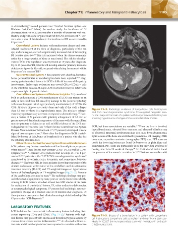
than 6.5 mm or there is a hypothalamic mass. The pituitary enlarge- Figure 71–2. Radiologic evidence of Langerhans cells histiocytosis
ment may spontaneously decrease or respond to chemotherapy. How- and CNS neurodegenerative syndrome. T2-weighted magnetic reso-
57
nance image of the brain of a patient with Langerhans cells histiocytosis
ever, a review of 22 patients with pituitary enlargement of 6.5 mm or showing hyperintense changes of the cerebellar white matter.
greater revealed that despite regression of the mass with therapy, all had
anterior pituitary deficiencies as well as MRI evidence of the CNS neu-
63
rodegenerative syndrome (see “Other Chronic Central Nervous System LCH, but these associations are variable. When the liver is involved
Disease Manifestations” below) and 17 (77 percent) developed clinical hypoalbuminemia, elevated liver enzymes, and elevated bilirubin may
58
signs of neurodegeneration. Most often the diagnosis of LCH is estab- be observed. Intestinal involvement may also cause hypoalbuminemia.
lished by biopsy of skin, bone, or lymph node of a patient who also has Lytic lesions of the bone are identified by plain films, CT imaging, MRI,
the pituitary abnormalities. bone scan, or positron emission tomography (PET) scan. PET scans are
Other Chronic Central Nervous System Disease Manifestations useful for detecting lesions not found by bone scan or plain films and
LCH patients may develop mass lesions of the choroid plexus, or gray or comparison PET scans are particularly good for providing evidence of
64
59
white matter. These lesions may contain CD1a+ DCs as well as CD8+ healing after 6 to 12 weeks of therapy. An institutional series found
60
lymphocytes. A chronic CNS problem that develops in 1 to 4 per- the presence of the somatic mutation in LCH lesions to correlate with
cent of LCH patients is the “LCH CNS neurodegenerative syndrome”
manifested by dysarthria, ataxia, dysmetria, and, sometimes, behavior
changes. 61,62 The brain MRI in these patients shows hyperintensity of the
dentate nucleus and white matter of the cerebellum on fluid-attenuated
inversion recovery (FLAIR) and T2-weighted images or hyperintense
lesions of the basal ganglia on T1-weighted images (Fig. 71–2). Atrophy
of the cerebellum also may be seen. The radiologic findings may pre-
59
cede the onset of symptoms by many years or can be found coincidently.
Among 83 LCH patients who had at least two MRI studies of the brain
for evaluation of craniofacial lesions, DI, other endocrine deficiencies,
or neuropsychological symptoms, 57 percent had radiologic neurode-
generative changes at a median time of 34 months after diagnosis. Of
these patients, one-quarter had clinical neurologic deficits develop 3 to
15 years after LCH diagnosis. 62
LABORATORY FEATURES
LCH is defined by characteristic inflammatory lesions including histi-
ocytes expressing CD1a and CD207 (Fig. 71–3). Patients with high- Figure 71–3. Biopsy of a bone lesion in a patient with Langerhans
1
risk disease may present with anemia and thrombocytopenia caused by cell histiocytosis. Langerhans cells cytoplasm and membrane stain pos-
marrow involvement and/or inflammation. 46,47 An elevated sedimenta- itively for CD207 (immunoperoxidase stain with hematoxylin and eosin
tion rate and thrombocytosis has been reported to correlate with active [H&E] counterstain).
Kaushansky_chapter 71_p1101-1120.indd 1105 9/17/15 3:50 PM