
Page 118 - Williams Hematology ( PDFDrive )
P. 118
94 Part II: The Organization of the Lymphohematopoietic Tissues Chapter 6: The Organization and Structure of Lymphoid Tissues 95
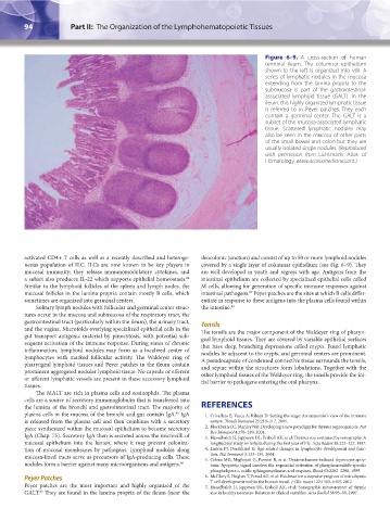
Figure 6–9. A cross-section of human
terminal ileum. The columnar epithelium
shown to the left is organized into villi. A
series of lymphatic nodules in the mucosa
extending from the lamina propria to the
submucosa is part of the gastrointestinal-
associated lymphoid tissue (GALT). In the
ileum, this highly organized lymphatic tissue
is referred to as Peyer patches. They each
contain a germinal center. The GALT is a
subset of the mucosa-associated lymphatic
tissue. Scattered lymphatic nodules may
also be seen in the mucosa of other parts
of the small bowel and colon but they are
usually isolated single nodules. (Reproduced
with permission from Lichtman’s Atlas of
Hematology, www.accessmedicine.com.)
activated CD4+ T cells as well as a recently described and heteroge- ileocolonic junction) and consist of up to 50 or more lymphoid nodules
neous population of ILC. ILCs are now known to be key players in covered by a single layer of columnar epithelium (see Fig. 6–9). They
mucosal immunity; they release immunomodulatory cytokines, and are well developed in youth and regress with age. Antigens from the
a subset also produces IL-22 which supports epithelial homeostasis. intestinal epithelium are collected by specialized epithelial cells called
84
Similar to the lymphoid follicles of the spleen and lymph nodes, the M cells, allowing for generation of specific immune responses against
mucosal follicles in the lamina propria contain mostly B cells, which intestinal pathogens. Peyer patches are the sites at which B cells differ-
87
sometimes are organized into germinal centers. entiate in response to these antigens into the plasma cells found within
Solitary lymph nodules with follicular and germinal center struc- the intestine. 88
tures occur in the mucosa and submucosa of the respiratory tract, the
gastrointestinal tract (particularly within the ileum), the urinary tract, Tonsils
and the vagina. Microfolds overlying specialized epithelial cells in the The tonsils are the major component of the Waldeyer ring of pharyn-
gut transport antigenic material by pinocytosis, with potential sub- geal lymphoid tissues. They are covered by variable epithelial surfaces
sequent activation of the immune response. During states of chronic that have deep, branching depressions called crypts. Fused lymphatic
inflammation, lymphoid nodules may form as a localized center of nodules lie adjacent to the crypts, and germinal centers are prominent.
lymphocytes with marked follicular activity. The Waldeyer ring of A pseudocapsule of condensed connective tissue surrounds the tonsils,
pharyngeal lymphoid tissues and Peyer patches in the ileum contain and septae within the structures form lobulations. Together with the
prominent aggregated nodular lymphoid tissue. No capsule or efferent other lymphoid tissues of the Waldeyer ring, the tonsils provide the ini-
or afferent lymphatic vessels are present in these accessory lymphoid tial barrier to pathogens entering the oral pharynx.
tissues.
The MALT are rich in plasma cells and eosinophils. The plasma
cells are a source of secretory immunoglobulin that is transferred into
the lumina of the bronchi and gastrointestinal tract. The majority of REFERENCES
plasma cells in the mucosa of the bronchi and gut contain IgA. IgA 1. Crivellato E, Vacca A, Ribatti D: Setting the stage: An anatomist’s view of the immune
85
is released from the plasma cell and then combines with a secretory system. Trends Immunol 25:210–217, 2004.
piece synthesized within the mucosal epithelium to become secretory 2. Blackburn CC, Manley NR: Developing a new paradigm for thymus organogenesis. Nat
Rev Immunol 4:278–289, 2004.
IgA (Chap. 75). Secretory IgA then is secreted across the microvilli of 3. Hasselbalch H, Jeppesen DL, Ersboll AK, et al: Thymus size evaluated by sonography. A
mucosal epithelium into the lumen, where it may prevent coloniza- longitudinal study on infants during the first year of life. Acta Radiol 38:222–227, 1997.
tion of mucosal membranes by pathogens. Lymphoid nodules along 4. Linton PJ, Dorshkind K: Age-related changes in lymphocyte development and func-
tion. Nat Immunol 5:133–139, 2004.
mucosa-lined tracts serve as precursors of IgA-producing cells. These 5. Cifone MG, Migliorati G, Parroni R, et al: Dexamethasone-induced thymocyte apop-
nodules form a barrier against many microorganisms and antigens. 86 tosis: Apoptotic signal involves the sequential activation of phosphoinositide-specific
phospholipase c, acidic sphingomyelinase, and caspases. Blood 93:2282–2296, 1999.
Peyer Patches 6. McClory S, Hughes T, Freud AG, et al: Evidence for a stepwise program of extrathymic
T cell development within the human tonsil. J Clin Invest 122:1403–1415, 2012.
Peyer patches are the most important and highly organized of the 7. Hasselbalch H, Jeppesen DL, Ersboll AK, et al: Sonographic measurement of thymic
83
GALT. They are found in the lamina propria of the ileum (near the size in healthy neonates. Relation to clinical variables. Acta Radiol 38:95–98, 1997.
Kaushansky_chapter 06_p0085-0096.indd 94 17/09/15 5:54 pm