Page 116 - Williams Hematology ( PDFDrive )
P. 116

92             Part II:  The Organization of the Lymphohematopoietic Tissues                                                                             Chapter 6:  The Organization and Structure of Lymphoid Tissues             93


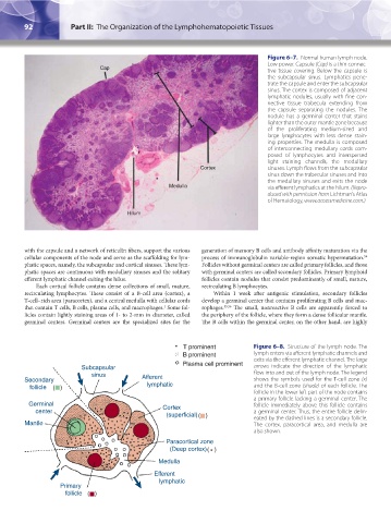
                                                                                          Figure 6–7.  Normal human lymph node.
                                                                                          Low power. Capsule (Cap) is a thin connec-
                                       Cap                                                tive tissue covering. Below the capsule is
                                                                                          the subcapsular sinus. Lymphatics pene-
                                                                                          trate the capsule and enter the subcapsular
                                                                                          sinus. The cortex is composed of adjacent
                                                                                          lymphatic  nodules,  usually with  fine con-
                                                                                          nective  tissue  trabecula  extending  from
                                                                                          the capsule separating the nodules.  The
                                                                                          nodule has a germinal center that stains
                                                                                          lighter than the outer mantle zone because
                                                                                          of  the  proliferating  medium-sized  and
                                                                                          large lymphocytes with  less dense stain-
                                                                                          ing properties. The medulla is composed
                                                                                          of  interconnecting  medullary  cords  com-
                                                                                          posed of lymphocytes and interspersed
                                                                                          light  staining  channels,  the  medullary
                                                                      Cortex              sinuses. Lymph flows from the subcapsular
                                                                                          sinus down the trabecular sinuses and into
                                                                                          the medullary sinuses and exits the node
                                                            Medulla                       via efferent lymphatics at the hilum. (Repro-
                                                                                          duced with permission from Lichtman’s Atlas
                                                                                          of Hematology, www.accessmedicine.com.)

                                               Hilum


               with the capsule and a network of reticulin fibers, support the various   generation of memory B cells and antibody affinity maturation via the
               cellular components of the node and serve as the scaffolding for lym-  process of immunoglobulin variable-region somatic hypermutation.
                                                                                                                        54
               phatic spaces, namely, the subcapsular and cortical sinuses. These lym-  Follicles without germinal centers are called primary follicles, and those
               phatic spaces are continuous with medullary sinuses and the solitary   with germinal centers are called secondary follicles. Primary lymphoid
               efferent lymphatic channel exiting the hilus.          follicles contain nodules that consist predominantly of small, mature,
                   Each cortical follicle contains dense collections of small, mature,   recirculating B lymphocytes.
               recirculating lymphocytes. These consist of a B-cell area (cortex), a   Within 1 week after antigenic stimulation, secondary follicles
               T-cell–rich area (paracortex), and a central medulla with cellular cords   develop a germinal center that contains proliferating B cells and mac-
               that contain T cells, B cells, plasma cells, and macrophages.  Some fol-  rophages. 55,56  The small, nonreactive B cells are apparently forced to
                                                          1
               licles contain lightly staining areas of 1- to 2-mm in diameter, called   the periphery of the follicle, where they form a dense follicular mantle.
               germinal centers. Germinal centers are the specialized sites for the   The B cells within the germinal center, on the other hand, are highly


                                                                T prominent           Figure 6–8.  Structure of the lymph node. The
                                                                B prominent           lymph enters via afferent lymphatic channels and
                                                                                      exits via the efferent lymphatic channel. The large
                                                                Plasma cell prominent
                                 Subcapsular                                          arrows indicate the direction of the lymphatic
                                    sinus           Afferent                          flow into and out of the lymph node. The legend
               Secondary                                                              shows the symbols used for the T-cell zone  (x)
                  follicle  ()                         lymphatic                      and the B-cell zone (shade) of each follicle. The
                                                                                      follicle in the lower left part of the node contains
                                                                                      a primary follicle lacking a germinal center. The
                 Germinal                                 Cortex                      follicle immediately above this follicle contains
                     center                                                           a germinal center. Thus, the entire follicle delin-
                                                            (superficial)()           eated by the dashed lines is a secondary follicle.
               Mantle                                                                 The cortex, paracortical area, and medulla are
                                                                                      also shown.
                                                           Paracortical zone
                                                             (Deep cortex)()
                                                         Medulla

                                                       Efferent
                                                          lymphatic
                          Primary
                             follicle ()


          Kaushansky_chapter 06_p0085-0096.indd   92                                                                    17/09/15   5:53 pm
   111   112   113   114   115   116   117   118   119   120   121