
Page 1206 - Williams Hematology ( PDFDrive )
P. 1206
1180 Part IX: Lymphocytes and Plasma Cells Chapter 76: Functions of T Lymphocytes: T-cell Receptors for Antigen 1181
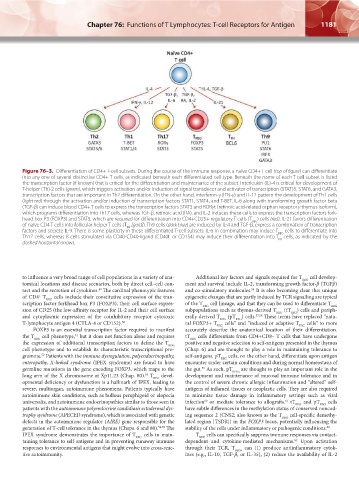
Figure 76–3. Differentiation of CD4+ T-cell subsets. During the course of the immune response, a naïve CD4+ T cell (top of figure) can differentiate
into any one of several distinctive CD4+ T cells, as indicated beneath each differentiated cell type. Beneath the name of each T-cell subset is listed
the transcription factor (if known) that is critical for the differentiation and maintenance of the subset. Interleukin (IL)-4 is critical for development of
T-helper (Th)-2 cells (green), which triggers activation and/or induction of signal transducer and activator of transcription (STAT)5, STAT6, and GATA3,
transcription factors that are important in Th2 differentiation. On the other hand, interferon-γ (IFN-γ) and IL-12 pattern the development of Th1 cells
(light red) through the activation and/or induction of transcription factors STAT1, STAT4, and T-BET. IL-6 along with transforming growth factor beta
(TGF-β) can induce blood CD4+ T cells to express the transcription factors STAT3 and RORγt (retinoic acid-related orphan receptor γ thymus isoform),
which programs differentiation into Th17 cells, whereas TGF-β, retinoic acid (RA), and IL-2 induces these cells to express the transcription factors fork-
head box P3 (FOXP3) and STAT5, which are required for differentiation into CD4+CD25+ regulatory T cells (T ) cells (red). IL-21 favors differentiation
REG
of naïve CD4 T cells into follicular helper T cells (T [gold]). Th9 cells (dark blue) are induced by IL-4 and TGF-β, express a combination of transcription
FH
factors and secrete IL-9. There is some plasticity in these differentiated T-cell subsets. IL-6 in combination may induce T cells to differentiate into
REG
Th17 cells, whereas B cells stimulated via CD40-CD40-ligand (CD40L or CD154) may induce their differentiation into T cells, as indicated by the
FH
dashed horizontal arrows.
to influence a very broad range of cell populations in a variety of ana- Additional key factors and signals required for T REG cell develop-
tomical locations and disease scenarios, both by direct cell–cell con- ment and survival include IL-2, transforming growth factor-β (TGFβ)
tact and the secretion of cytokines. The cardinal phenotypic features and co-stimulatory molecules. It is also becoming clear that unique
49
56
of CD4 T REG cells include their constitutive expression of the tran- epigenetic changes that are partly induced by TCR signalling are typical
+
scription factor forkhead box P3 (FOXP3), their cell surface expres- of the T REG cell lineage, and that they can be used to differentiate T REG
sion of CD25 (the low-affinity receptor for IL-2 and their cell surface subpopulations such as thymus-derived T REG (tT REG ) cells and periph-
and cytoplasmic expression of the coinhibitory receptor cytotoxic erally derived T REG (pT REG ) cells. 57,58 These terms have replaced “natu-
T-lymphocyte antigen 4 (CTLA-4 or CD152). 50 ral FOXP3+ T REG cells” and “induced or adaptive T REG cells” to more
FOXP3 is an essential transcription factor required to manifest accurately describe the anatomical location of their differentiation.
the T REG cell phenotype, but it does not function alone and requires tT REG cells differentiate from CD4+CD8– T cells that have undergone
51
the expression of additional transcription factors to define the T REG positive and negative selection to self-antigens presented in the thymus
cell phenotype and to establish its characteristic transcriptional pro- (Chap. 6) and are thought to play a role in maintaining tolerance to
gramme. Patients with the immune dysregulation, polyendocrinopathy, self-antigens. pT REG cells, on the other hand, differentiate upon antigen
52
enteropathy, X-linked syndrome (IPEX syndrome) are found to have encounter under certain conditions and during normal homeostasis of
germline mutations in the gene encoding FOXP3, which maps to the the gut. As such, pT REGs are thought to play an important role in the
59
long arm of the X chromosome at Xp11.23 (Chap. 80). T REG devel- development and maintenance of mucosal immune tolerance and in
53
opmental deficiency or dysfunction is a hallmark of IPEX, leading to the control of severe chronic allergic inflammation and “altered” self-
severe, multiorgan, autoimmune phenomena. Patients typically have antigens of inflamed tissues or neoplastic cells. They are also required
autoimmune skin conditions, such as bullous pemphigoid or alopecia to minimize tissue damage in inflammatory settings such as viral
universalis, and autoimmune endocrinopathies similar to those seen in infection or mediate tolerance to allografts. tT REG and pT REG cells
60
61
patients with the autoimmune polyendocrine candidiasis ectodermal dys- have subtle differences in the methylation status of conserved noncod-
trophy syndrome (APECED syndrome), which is associated with genetic ing sequence 2 (CNS2; also known as the T REG cell-specific demethy-
defects in the autoimmune regulator (AIRE) gene responsible for the lated region [TSDR]) in the FOXP3 locus, potentially influencing the
generation of T-cell tolerance in the thymus (Chaps. 6 and 80). 54,55 The stability of the cells under inflammatory or pathogenic conditions. 62
IPEX syndrome demonstrates the importance of T REG cells in main- T REG cells can specifically suppress immune responses via contact-
taining tolerance to self-antigens and in preventing runaway immune dependent and cytokine-mediated mechanisms. Upon activation
63
responses to environmental antigens that might evolve into cross-reac- through their TCR, T REGs can (1) produce antiinflammatory cytok-
tive autoimmunity. ines (e.g., IL-10, TGF-β, or IL-35), (2) reduce the availability of IL-2
Kaushansky_chapter 76_p1175-1188.indd 1181 9/17/15 4:01 PM